
Lab investigations are a crucial part of research and diagnosis. The gross and microscopic study of tissue helps understand the normal and the abnormal. Histology and pathology are two such branches of science that study any given structure under the microscope to help the examiner understand what is going on at a microscopic level.

So what is the difference between them?
Histology is the field of science where tissue is studied under the microscope to see the microscopic composition of that tissue. Pathology is the field of study where the disease process is studied. Pathology includes examining the tissues grossly (physical changes) and histologically.
Histological study of any tissue enables the examiner to see the various layers of the tissue and the different types of cells forming the tissue. A histological examination is done to study the normal microscopic structure of the tissue and to see the changes that occur in the tissue during any disease process.
Pathology is a broader subject and includes studying the physical and histological changes in the tissue to understand the disease process. The study of microscopic changes in any pathology is known as histopathology. Pathologists also perform various other tests like immunohistochemistry and molecular tests like FISH (fluorescence in situ hybridization). These tests help them study the disease better, along with histological exams.
What is Histology?
Histology involves the microscopic study of any organ or tissue. It can be done for research purposes or to study a disease process in a pathology lab.
An example of a histological study is the biopsy— in which a tissue is taken out and sliced into thin sections. These thin sections are then placed on a slide for staining and visualized under the microscope. In the histological study of the biopsy sample, the different layers of the tissue with the pathological changes in them can be seen.
Histology can be done to study both the normal structures and the abnormal changes in the tissue.
Also, histology should not be confused with cytology, which is the study of cells.
In cytology, cells are studied under a microscope after preparing a smear of the cells and staining them with appropriate stains. The examples of cytological study are:
- FNAC (fine needle aspiration cytology), which is done for breast lumps and suspected thyroid cancers, and
- Pap smear– done to screen for cervical cancer.
Only cells are visualized in this study, not the layers of the tissue. If there are any pathological changes in the cells, your doctor may proceed to the next test, i.e. biopsy.
The histological study is more helpful in giving a confirmatory diagnosis. Cytology helps hint at pathology.
The cytological study is, however, less invasive and is therefore sometimes done first to see if there is an abnormality present or not. On the other hand, a biopsy is more invasive as it requires a chunk of tissue that can be obtained in a clinic with or without local anesthesia or may need surgery to get the sample– depending on the condition.
You can follow the following youtube video to see the histological view of a few different types of cells in the human body:
What is Pathology?
Pathology is a branch of medicine that studies disease processes. It includes the study of how various diseases cause changes in a particular organ, tissue, or cells and what happens because of these changes.
The biopsy is collected by the physician, surgeon, or pathologist and sent to the pathology lab for study. Slides are prepared using these samples for the assessment by the pathologist. A pathological study helps make a confirmatory diagnosis in many conditions.
A pathological examination will include the gross exam of any organ or tissue and the changes happening at a microscopic level. These changes may vary at different stages of the disease.
After studying the microscopic changes, a pathologist comes to a diagnosis of the disease. The study will also help in staging the disease (wherever applicable).
Histology, cytology, and other advanced tests like molecular tests, etc, are a part of pathological exams as all of them help the pathologist understand the disease better.
Pathological studies are essential in hospital settings to make a diagnosis and in the research sector to understand the pathological processes of new diseases.
You can see the following video to understand what a histopathologist does:
Career as a Pathologist
A pathologist is a medical doctor that specializes in pathology. They play an essential role in making a diagnosis of various illnesses. Based on the pathological findings combined with the symptoms and other findings, a diagnosis is made. Your primary physician or surgeon then plans the treatment based on the combined information.
To become a pathologist, you need to go through medical school to get your primary medical education and then pursue a residency/specialization in pathology.
Career as a Histotechnician
A histotechnician helps prepare the slides for the microscopic examination in a lab. They slice, fix, stain, and mount the specimen on a slide. The pathologist then examines this slide.
A good slide preparation is the most crucial step in a histopathological examination. Therefore, a skilled histological technician is a vital part of any lab.
A histotechnologist also performs a similar role in a lab, but their training is more advanced when compared to that of a histology technician. They learn more about the background science involved in their work. Histotechnologists are more likely to get promoted to supervisor posts further in their career and may get the responsibility to oversee others’ work in the lab.
A histotechnician can later do the certificate program to become a histotechnologist if they wish to learn more or if they desire to play more of a leadership role in the future.
It is a good option for those who don’t want to be a doctor or a nurse (i.e. those who don’t want to deal with the patients directly) but still want to be a part of medicine in some way.
There are separate certificate programs to become a histotechnician and a histotechnologist. The duration of the course varies, with the duration for the latter being longer and more advanced.
Career Opportunity in a Pathology Lab
The demand for a skilled pathologist and a skilled histotechnician is always there. Both of their skills determine an accurate diagnosis. With more and more research going on, pathologists and histotechnicians can work in a hospital, clinic, or research.
A skilled technician is a valuable addition to any lab as their skillful preparation of the slides is the first step to any diagnosis and the smooth functioning of the lab.
On the other hand, the skill of a pathologist determines accurate diagnosis in many conditions. They help physicians and surgeons see what is happening at a microscopic level in any disease. Based on their findings, the treatment is planned, and the prognosis is understood. They are essential for medical diagnosis in all the clinical branches of medicine.
With new research going on in the field of medicine– good pathologists and histotechnicians are required. They both play a crucial role in research and diagnosis.
Histology Vs Pathology: Points to Remember
So, what you need to remember is:
- Histology is the microscopic study of a tissue, which enables the examiner to understand the normal microscopic composition and the abnormal microscopic changes in tissue.
- Pathology is a broader field of medical science that focuses on studying the disease process and may use histological studies to understand the disease better.
- Both of them are promising career options.
You can read about how to prepare a microscope slide mounting medium here: How To Make Microscope Slide Mounting Medium?
3D Animation of Human Excretory System and Kidney Anatomy and Function
In this 3D animation we go over the anatomy and physiology of the human excretory system, the kidneys, nephrons, and glomeruli.
This 3D animation of the human excretory system describes in detailed yet easy to understand language the anatomy and physiology of the kidneys, nephrons, glomerulus, and the processes of excretion, filtration, reabsorption and secretion, showing the location and anatomy of all the important structures involved, including the Bowman’s capsule, loop of Henle, pyramids and collecting ducts. USMLE test content.
Review:
The kidneys are the primary organs involved in the process of excretion. The kidneys are part of the urinary system.
The two kidneys on either side of the backbone are connected to two ureters that empty into the urinary bladder down below. The urethra is the tube at the bottom of the urinary bladder. Kidneys are bean shaped, dark red, somewhat flattened and located under the diaphragm near the back wall of the abdominal cavity. Each kidney weighs about 150 grams and is 10 cm long. The left kidney is slightly higher than the right kidney.
Kidneys filter metabolic waste out of blood and remove water. This allows the urinary system to regulate composition of fluids including salts, water, waste. The kidneys also regulate blood pressure, maintain pH. Kidneys process more than 200 liters of blood a day.
A kidney consists of:
renal sinus- cavity where you can find fat, nerves, blood vessels and renal pelvis
renal pelvis- funnel shape part of ureter that funnels urine flowing into kidney
Ureter- long tubes that move urine to urinary bladder from kidneys
outer renal Cortex- outer portion of kidney between medulla and renal capsule
inner Medulla- innermost part of kidney, divided into renal pyramids,
renal pyramids- cone shaped structures of kidney that have a striped appearance due to being made up of the nephrons Loops of Henle
A Nephron is the functional unit of kidney. Name comes from greek Nephros which stands for kidney. Nephrons are very small filters (millions tightly packed). A part of the nephron is in the cortex of kidney and part is in medulla. Nephrons are about 4cm long. The renal corpuscle is made up of cluster of a capillaries named the glomerulus and a sac like structure called the Bowmans capsule around the glomerulus. The renal tubule, the other part of the nephron that starts at the Bowmans capsule and ends at ducts of kidney is made up of 3 parts:
- proximal convoluted tubule- part by bowmans capsule thats why called proximal (in cortex of kidney)
- loop of henle- in renal medulla, and descending and then ascending limb
- distal convoluted tubule- named because far from bowmans capsule, that one empties into the collecting duct (located in cortex of kidney)
The collecting duct is where many nephrons drain into (it is in medulla of kidney). Many
collecting ducts drain into the renal pelvis which drains into the ureter.
Thin walled capillaries run the entire length of the nephron. They bring in blood with metabolic waste.
Renal artery and vein:
The renal artery (branch of aorta) brings in unfiltered blood into kidney; filtered blood leaves via the renal vein.
An afferent vessel brings blood into the glomerulus. An efferent vessel takes blood out of glomerulus. Glomerulus comes from latin Glomeris meaning ball of yarn. The glomerulus is a ball of capillaries between afferent and efferent vessel. The ball increases surface area for filtration into the bowmans capsule.
Waste in the form of urine passes from the kidneys down the ureters into the urinary bladder where it is stored until it can be released. Pressure in the bladder from stored urine causes an urgency to release. Urine is formed through filtration, reabsorption, secretion, excretion. Items not filtered include proteins and RBCs.
Waste (uric acid, urea-nitrogenous waste) is removed from blood in the glomerulus. Urine is formed in the nephron from the glomerular filtrate.
Reabsorption occurs of some substances such as salts, excess water, amino acids, glucose that are filtered together with nitrogenous waste that are needed by body. Reabsorption occurs along proximal convoluted tubule of nephron into peritubular capillaries and back into circulation. More reabsorption happens in the loop of henle for things such as magnesium, calcium, potassium, sodium. This is made possible by aquaporins
Secretion occurs when reabsorbed waste is secreted into blood from urine flowing through the tubes of the nephron. Secretion occurs when waste that did not get filtered goes from capillaries into proximal and distal convoluted tubule and becomes urine. Substances secreted include urea, creatine, uric acid, hydrogen, ammonium, potassium ions
Click here for an animation on human brain anatomy and physiology.
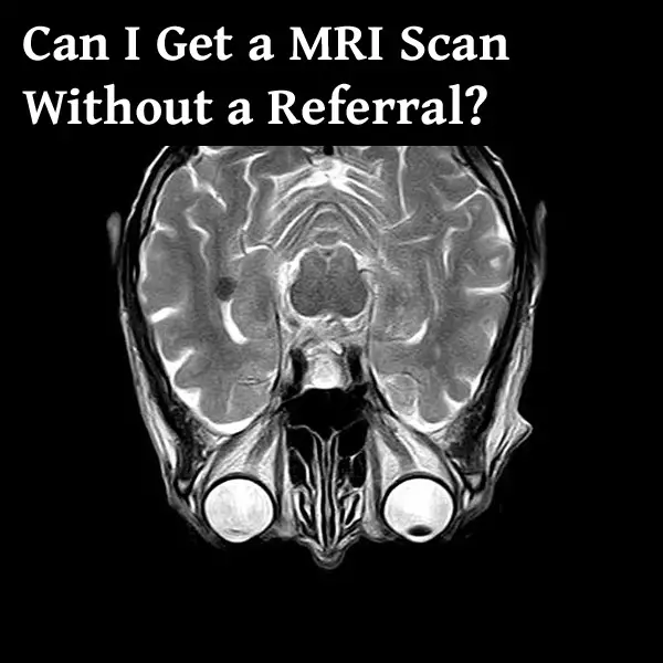
Can I Get an MRI Scan Without a Referral?
Thanks to capitalism, you can get a lot of things without asking for much permission or getting a referral. This includes many diagnostics like an MRI (Magnetic Resonance Imaging). An MRI can be a very expensive diagnostics procedure. It is also very involved and takes a bit of time to set up.
The MRI machine is also quite large and expensive so every clinic is not equipped with one. In fact, many small clinics will refer you to larger hospitals. However, there are some instances where a person may want to get an MRI without the referral of a primary care physician. This could be for a number of reasons including general curiosity, getting a second opinion, or even preparing for a malpractice lawsuit.
Because MRI is of the more expensive and higher quality scans, it can be more difficult to get. These machines are also not available everywhere, so you have to take into account travel and accessibility. The point is that it can be done.
Regardless of the reason for getting an MRI without a referral, it is possible. It may require a bit more effort and definitely more money. Many insurance companies will only pay for what your physician will prescribe. Anything outside of that will be personal costs unless you have an HSA (Health Savings Account) that you can access.
What is an MRI?
An MRI is a non-invasive imaging technology that is used to view anatomical structures within the body. This is done through the use of magnetic resonance. This technology can be used to diagnose injury and disease as well as monitor the body during treatment. MRI’s are usually taken for specific regions of the body. For example, a common MRI is for the brain and spinal cord. This is a great way to check for bleeding, swelling of the brain and/or around the spinal cord. It is also a great way to check for damage.
Another common region is the abdominal area. This is where doctors will normally check for internal injuries or other issues like tumors. An MRI can check for damaged organs like the spleen which is a typical site for internal bleeding.
An MRI works by aligning the positively charged particles (protons) within the body to a magnetic field. Radiofrequency waves are pulsed through the body. As those waves interact with those protons, it results in images that allow physicians to decipher between specific tissues and organs.
This procedure causes no harm to the patient. However, it can be disconcerting during the process. The patient has to lie very still while inside of a hollow tube. For those with claustrophobia or any other anxieties, make sure to speak with your physician about medical intervention.
Why Do Doctors Refuse MRI?
MRIs are usually prescribed by a primary care physician or an emergency doctor if there is a suspicion of injury or disease. These are very expensive machines and require specialized technicians to run them. In areas that do not have immediate access to these machines, it can be difficult to schedule a scan. In addition to that, for non emergent scans, there may be a list of those waiting for the machine.
Doctors may refuse to prescribe an MRI if they don’t see a viable reason to do so. It could be that a patient may not be showing signs of internal injury or disease. In cases of needing a neurological MRI, a full neurological exam should be performed prior. In these cases an MRI would be used to confirm any disease based on neurological defects.
In the case of an abdominal MRI, a physical exam should be performed prior as well. If there is no tenderness, distension, or tightness of the abdomen, a physician may not readily prescribe an MRI to a patient even if they have suffered physical trauma.
That is not to say that there aren’t other options. Some of those options are also cheaper than an MRI. An X-ray can reveal a lot about what is going on within the body. Even when it comes to organs. Ultrasound is also a fast way to see what is going on inside of the body as well. Ultrasound machines are also becoming more readily available in hospitals. There are even some technologies where you can attach a probe to a smart device like a phone or tablet and perform an ultrasound at the bedside.
There could be many reasons why your doctor did not prescribe an MRI in a specific incident. However, if you disagree with his/her/their recommendations, you can go around that.
How to get an MRI scan without a Referral
Getting an MRI without a referral can be a little complicated. There aren’t many hospitals that will do it without a physical exam and some sort of hospital record. Fortunately, you can get access to your own records and have those transferred to a different hospital and start there.
You may have better luck at privately owned hospitals or at free-standing imaging facilities. These hospitals are not as bound by budgetary needs as a more publicly-funded hospital. Also, as medical costs increase, people are shopping around for these types of medical procedures. There are locations outside of hospitals that only do MRI scans or any other diagnostic scans a la carte.
It is best to call around and see what these locations offer and what is required of you before arriving. Make sure to ask about payment options as well as talking with a technician or an actual physician afterwards to interpret your results. The average person will not know what they are looking at if they were to see an MRI scan on a screen. You are going to need to talk with a professional about what is there.
If there is anything abnormal, the doctor will be able to give recommendations for next steps, further referrals, etcetera. If there are no present concerns, you can speak with another physician about next steps as well.
How Much Does an MRI Scan Cost?
The cost of an MRI can vary based on a number of factors including location. It could be based on what area(s) need to be scanned as well. There are also different types of MRIs (closed/open). Generally, MRIs range from $400 to $3,500. This could also change depending on if you are using insurance. Without insurance, an MRI could cost closer to $5,000.
If you are going to a free-standing facility, these prices may be lower than what you will find in a hospital. Insured and uninsured patients make use of these spaces. If you are going to get an MRI without a referral, your insurance may not pay for this testing. That is definitely going to be a major consideration. Depending on who you open the account with, you could use an HSA (health savings account) to pay for this service.
Possibly the better option would be to seek out a second opinion and request a referral for an MRI scan from your second physician. This could eliminate many of the obstacles you may run into. While your reasons for seeking out a private MRI scan may be valid, it could also be more difficult than if you go through the usual channels.
Proceeding from Here
In conclusion, an MRI is a high-tech method of looking at the anatomical structures within the body like the brain, spinal cord, and abdominal organs. While there are other technologies that could be used during an examination, an MRI is definitely more advanced. Some doctors may have many reasons for not prescribing an MRI, but if you disagree, there are ways around that. You can seek out a private MRI scan. It may be more difficult and definitely more expensive because your insurance is less likely to pay for it. Due to the increase in medical costs, there are more options for people to shop around with or without insurance.
You will at some point need the help of a professional after getting your scans. A professional will help you interpret those scans and determine next steps. While it is not recommended to get any type of diagnostic procedure without a prescription or a referral, many are available to people without them. You just have to work a bit harder for it.
Click the following link to learn the difference between radiology and medical imaging.
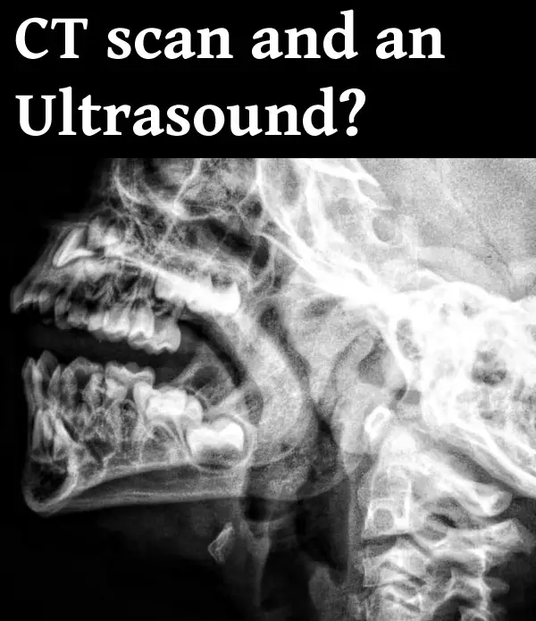
Why Would you Need a CT Scan and an Ultrasound?
With the advancement in technology, there are so many new technological advances that medical science has seen in the past few decades. Many imaging modalities are available to choose from now. Some investigations are more sophisticated than others, but each of them has its unique application. Two such imaging modalities that are very commonly used are the USG and CT scan.
So, what’s the difference between them, and when are an ultrasound and a CT scan needed?
An Ultrasound uses high-frequency sound waves to produce an image. It has certain limitations to what structures it can see. It is usually done to scan soft organs and structures. A CT scan is done using X-rays and it produces more detailed images. It can be used to see both soft tissues and bones. Combined, a CT scan and an ultrasound provide a full picture of the condition, including soft structures and bones, where an ultrasound is more readily available and affordable.
A CT scan is more sophisticated and produces more detailed images. It forms multiple layers of images of the organ under study from all directions (360 degrees). This allows the medical professionals to see exactly where the lesion or defect is and what is their extent or severity. This helps in planning for better treatment– medical or surgical. They can be used to look at tumors, internal injuries, and other conditions located anywhere in the body. They are great at detecting bleeding and are also great at detecting renal stones, which are often not seen in USG and X-rays. They may also be combined with other sophisticated investigations like MRI to get an even better idea regarding the condition under study. In addition to internal organs, they are also used to look at bones and related conditions, including very fine fractures.
Ultrasound on the other hand is most commonly used to visualise abdominal organs and other soft structures. Conditions like appendicitis, pancreatitis, hepatomegaly, splenomegaly, PCOS, abdominal pain, etc are some of the most common conditions diagnosed with the help of a USG. Another most important use of USG is the antenatal scanning during pregnancy, to monitor the development of the fetus. USG has limited application when compared to a CT scan but they are more commonly available, affordable, radiation-free, and good enough for diagnosing many conditions.
More applications of ultrasound and CT scans are discussed below.
What Is An Ultrasound?
Also known as sonography, USG is an imaging modality that uses sound waves to produce images of organs and other structures in the body.
An ultrasound sends a sound wave towards the organ under study and produces an image based on how long it takes for the sound wave to reflect— much like sonar.
Since it doesn’t use radiation to create an image, it’s a very safe, non-invasive radiological investigation. Ultrasound is a very commonly used investigation, and is usually the very first radiological investigation doctors order in a wide number of cases because it’s easily available, is more affordable as compared to CT scan and MRI, and is good enough for making certain diagnoses.
Where Is An Ultrasound Used?
Ultrasound has a wide range of applications. It can be used to investigate the abdomen, the blood vessels, and the reproductive tract– to name a few.
Different organs may require a different mode of sonography for a better examination.
Transabdominal Sonography (TAS):
This is the most commonly done USG, which— as the name suggests— is used to look for abdominal organs. This mode of sonography is used to examine the GI tract, kidneys ureter, bladder, pancreas, liver, uterus, Fallopian tubes, etc— to name a few.
So, the application of TAS is very wide. Some of the conditions where your doctor may ask for a TAS are:
- Abdominal masses
- Renal issues, like pyelonephritis, recurrent UTI, hematuria, etc
- Ascites
- Splenomegaly
- Hepatomegaly
- Uterine conditions like infections, developmental anomalies, etc
- Infertility— initial investigation
- Pregnancy— to monitor the development of the foetus
- Pancreatitis
- LIver and spleen injuries in the cases of trauma, in the emergency setting.
- Abdominal pain, etc
Transvaginal Sonography (TVS)
This type of ultrasound is done specifically to see the uterus, Fallopian tubes, ovaries, and even bladder. The USG probe is inserted via the vagina and the imagining is performed.
It can be performed in conditions like
- ectopic bleeding,
- ensure the placement of an IUD,
- infertility,
- uterine fibroids,
- abnormal vaginal bleed,
- to look for placental abnormalities,
- diagnosis of very early pregnancy,
- miscarriage.
Transrectal Ultrasound (TRUS)
In TRUS the probe is inserted into the rectum. Also known as endorectal ultrasound, it is used to look at the rectum and surrounding structures. It is also used to assess prostate and to perform biopsies.
Transesophageal Echocardiography (TEE)
TEE is done to examine the structure of the heart and the arteries connected directly to the heart. The probe is inserted via the mouth into the oesophagus to perform TEE.
Ultrasound can also be done to examine the blood flow, joint inflammation, issues of the eyes, pericardial effusion, pleural effusion, breast lump, and to perform various guided procedures like ultrasound-guided biopsy, etc.
What Is A CT Scan?
A CT scan is a radiological investigation that utilises X-rays to produce images of the internal organs and gives a much more detailed image of bones, muscles, blood vessels, and various organs.
The X-rays are beamed from different directions and different levels, and this helps in the formation of various views of the same organ with much more detail. The image collected at the X-ray plate is sent to a computer where the software produces a 2D image of the structure under study. With the advancement in technology, 3D images are also available now.
It can be done with contrast (to visualise blood vessels better), or without contrast (which is done regularly). Some people may be allergic to this dye, and the severity of the hypersensitivity can vary from person to person. It is important to pass on this information to the doctor if you’ve had a prior reaction to any of the dyes,
A CT scan may pose some risk to pregnant women. It is usually only ordered during pregnancy in conditions where they can’t wait until after the delivery to perform the scan, and when it’s essential in addition to USG and MRI. The treating doctor or the radiologist will make the patient aware of the associated risks if any. Although, the dose of the X-rays used for CT scans is adjusted to a much lower value for pregnant women, and when necessary, CT scans can be done during pregnancy.
Where Is A CT Scan Used?
CT scans can be done in a wide number of cases as well. It’s one of the very commonly used imaging modalities in the emergency department, as it gives a very detailed image of the internal organs and takes much less time to scan the body when compared to an MRI.
A CT scan can be done for:
- Cases of head injury. Various types of intracranial haemorrhage can be visualised in a CT scan.
- Stroke
- Seizures
- Brain tumour
- Chronic headache
- Hydrocephalus
- Congenital intracranial conditions
- Ear, nose, and throat conditions
- Vascular malformations
- Ocular and orbital lesions
- Facial fractures
- Spine injury
- Renal stones
- COVID 19
- To look for lung diseases
- To look for abdominal diseases
- Abdominal injuries and haemorrhages
- To look for metastasis
- Like USG and MRI, it can also be used to perform various guided procedures, like biopsies.
These are some of the conditions where your doctor may ask for a CT scan.
So, What You Need To Remember About a USG and a CT scan?
CT scan and ultrasound are two different types of modalities used for medical diagnoses. USG uses high-frequency sound waves to create an image, while CT scan uses X-rays to produce the image of internal organs.
CT scan gives a more detailed image when compared to a USG, but both have their own set of applications. Even though a CT scan gives a superior image, it’s not required to be done in many cases— where a USG image is enough to make a diagnosis.
Here is a useful video that shows more details about CT scans:
To learn more about radiological imaging, follow this link: Medical Imaging Vs Radiology: What’s The Difference?
Is 3D Modeling Harder than Drawing?
3d models are used in many industries nowadays including medical illustration and animation. If you are interested in taking up 3d modeling as your career, there are many opportunities. If you are good at creating drawings, you might be interested in exploring 3d modeling. Now, there will be many questions that come to your mind such as what types of drawings are needed for a 3d model?

Lets discuss how 3d modeling is different than drawing, how they are similar and which is easier.
3d modeling is not harder than drawing in the sense that the computer assists you with automated lighting, texture application, and perspective changes. 3d modeling is not easy however, as it requires good knowledge of the tools in your modeling software, which are only sometimes intuitive like clay sculpting. Modeling software often has a steep learning curve.
Do I need to be good at drawing to be a 3D modeler?
When it comes to drawing, it is a 2-dimensional flat medium. You can put it on paper. You can easily hold the paper in your hand. The drawing represents the figure in an X-Y plane, whereas 3D is representation in the X-Y-Z plane.
There are different types of 3d modeling out there, such as polygon modeling, NURBS, subdivision surfaces, etc. All these types of 3d modeling do not demand great drawing skills. But, there are a few advantages that you can get if you are good at drawing. For example, you will have a better understanding of perspective if your drawing skills are really good. Also, it helps to improve the lighting on your 3d models. So, overall your drawing skills can help to enhance 3d modeling skills. However, there will be only a small advantage.
If you are new to 3d modeling and not good at drawing, it will take only a couple of months to learn the modeling software. You can quickly learn all the techniques to create good 3d models with the help of many tutorials available on the internet. Once you are good at using the software, your drawing skills may not be that important.
3D modeling by sculpting with a pen and tablet
There are a lot of discussions going on amongst both professionals as well as amateur 3d modeling artists about whether a pen and tablet are needed for modeling a 3d object. If you use a mouse instead of a pen for sculpting the 3d model in such programs as Zbrush or Maya, you will not get good control. A pen will give you better control over each stroke. This type of 3d modeling is where good coordination and being used to drawing comes in handy. However, you dont need to have drawing skills such as shading, since it is sculpting.

When you use a mouse for sculpting, you need to press and hold your mouse and move it in different directions. You will be putting lots of pressure on your fingers, palm, and hand by doing this activity. If you continue it for a long time, you will get hurt.
When you sculpt using a pen and tablet, movement will be along with the hand and the shoulders. So, you will be more comfortable using a pen and tablet instead of a mouse.
Types of drawing required for 3d modeling
Now, we will look at the different types of drawings that are needed to create a 3d model.
1. Texture
It is the texture that brings the 3d model to life. You will have to use many textures to complete a 3d object. For example, if you are 3d modeling a person, you will have to get textures for the person’s face, different clothing, shoes, etc.
2. Basic 2D Shapes
Most of the 3d modeling software will need some 2D shapes to start the modeling process. The shape can be a simple one like a square, rectangle, circle, etc. It is pretty easy to draw such shapes. Sometimes, the shapes can be complex ones also. You can create complex shapes inside the modeling tool itself. Once the modeling software gets the shape, you can extrude the shape in the z-axis to make it a 3d shape. Again, no special drawing skills required.
3. Drawings from Different Points of View
If you are creating a 3d shape for industrial or medical parts, you need to be very specific about the part’s dimensions. Here, you need to create the reference drawings of the part first with appropriate dimensions. You will have to create drawings with different angles of view. It includes a front view, side view, top view, and bottom view. Once you create these drawings, you can feed them to the 3d modeling software. The software will create the 3d model taking these different angles of view as the input.
3d Modeling Vs Drawing
We will compare 3d modeling with drawing. It will get you a better understanding of the differences between the two.
1. Lighting
When you create a 2D drawing, you need to simulate lighting different portions of the image according to the lighting conditions that exist in your imagined picture. Some parts of the image will be under the shadows, whereas others will be in the light. It requires some skill and will come only through practice and observations such as those in real life.
In the case of a 3d model, lighting is relatively easy. First, you need to place the light source at an appropriate distance and angle from the subject. Then, you can easily see the lights and shadows created by the light source on the subject.
But, lighting plays a very important role in creating 3d art. So, it is essential to know where to place the light source (sun or artificial lights in the 3d software). Otherwise, you won’t be able to give the required feel to the 3d art, especially if going for realism.
If you are doubtful regarding the placement of the light source in a 3d software program, you can move it around and see the effect. It will help you to get a better idea. But this option is not available for a 2d image. You need to have a better pre-visualization in the case of a 2d image, such as with real life objects.
2. Perspective Learning
Perspective comes into the picture for both 3d and drawings. In the case of drawings, you need to have a better understanding of perspectives. Only then you will get the desired results in the drawing. On the other hand, you don’t have to overthink about perspective for creating a 3d model. You just need to enter the actual measurements for the model, scene camera settings such as field of view, focal length. Then, the camera in the 3d modeler will take care of the perspective part.
3. Creating the texture
If you want to create a texture in 2D, it will take a lot of work from scratch. For example, while drawing a wooden block in 2d, you need to give it appropriate texture to make it look like wood. It will take a good amount of time. You must also consider the light falling onto the subject while drawing the texture.
When you create 3d models, it is pretty easy to add a texture. You can apply textures for wood, metal, skin, etc. You can apply existing materials. You can modify them. You don’t have to think about lighting here. You can input the desired texture in 2d form to the 3d modeler software, and it will apply the texture to the model. This can be a photographed texture, a drawn one, or a combination. You need to know things like how to blend textures at edges, how to repeat them etc.
4. Fixing Mistakes
You will normally be making lots of mistakes when you create a 2d drawing or a 3d model. The point here is how easy it is to fix your mistakes in both cases.
If you consider the case of a 3d model of a person, you can change the clothes and lights separately in the model. If you make any mistake in clothes or other parts of the model, you might have to resculpt, remodel the polygons, or go back to the original curves used to make the model. This can be time consuming but the software may have the later steps already saved in modeling history so that all you may need to change is the original curves.
In the case of a 2d drawing, fixing a mistake can be really tough, especially if the medium does not allow erasing. Even if you draw on a tablet with layers, it will take some time.
5. Getting feedback through the creation process
It is always good to get feedback from the tool during the creation process. It will help in both 3d as well as 2d. This feedback mechanism is available for 3d modeling. The 3d modeler software will allow you to see the texture applied as well as the light during the modeling process. It will help you get a better idea of how your 3d model will look. Thus, it allows you to make the necessary changes in your workflow to improve your model. There is no need to wait to see the final finished model to see how it looks. It helps to fix the mistakes and improve your 3d model.
In the case of drawing, you will be able to figure out how it will look only in the final phase of its creation. So, you need to be more precise and accurate when you create a drawing. There is no scope for fixing errors during the initial creation phase. It makes the drawing really tough compared to the 3d modeling part.
6. Segmented approach
The 3d software programs follow a segmented approach for creating 3d models. The three main components of both 3d as well as 2d drawings are color, composition, and lighting. The segmented approach in 3d allows you to isolate each of these parameters and try them on your design to see the impact, for example you can move objects around in your scene freely to change composition.
When you take the case of a drawing, all these three parameters work together. You cannot isolate one and go ahead with your creation.
Here is an interesting video on Blender 3D software and drawing:
Consider reading this article on which is better for modeling, Maya or 3Ds Max.
Can Urgent Care do MRI? Norms and Exceptions
Urgent care facilities play an important role in tending to the non-life-threatening cases outside of the emergency department for which the patient would have otherwise gone to the emergency department. They provide their services at a lower price than any typical emergency department, and anyone can walk in anytime to get the treatment that they need as they don’t need referrals.

However, the services they provide are limited, and so are the investigations they offer. So, can you get an MRI done in an urgent care facility?
Urgent care facilities usually don’t offer MRI scans. However, some urgent care facilities do. A typical urgent care facility offers basic investigations like X-ray, ECG, USG, blood work, etc. The services they offer may vary. Calling in beforehand will help you find out what services you can get.
MRI or magnetic resonance imaging is a radiological imaging method that utilizes super magnets to produce radiological images of the area under study. It’s a very sophisticated and safe imaging modality, as it doesn’t utilize radiation to scan the body, unlike CT scan. Even though MRI scans play an important role in making a clinical diagnosis in modern medicine, their availability is often limited, and so is their application, especially in emergency situations. You will most likely find them in larger hospitals or health care facilities, but they are usually not available in urgent care facilities.
What is an urgent care facility? When is MRI needed on an urgent basis? We’ll explore these questions and more in this article.
What Do Urgent Care Facilities Do?
Urgent care facilities provide basic medical facilities, like investigations and treatment, for non-emergency conditions. They also provide other medical services like vaccinations, immunizations, and certain screening tests.
Urgent care facilities are a great place to seek help for non-life-threatening conditions, as they don’t need a referral to see a patient and are cheaper when compared to any emergency department. The waiting time in urgent care facilities is also usually shorter. In addition to this, they help reduce the load on the emergency services by taking on the cases that can be dealt with outside of the emergency department.
It is, however, essential that the patient understands that urgent care facilities don’t offer emergency investigations or interventions. They may even lack many superior investigations and scans, like an MRI, etc. Therefore, if there is any health emergency, you should only go to the nearest emergency department, or call 911.
What Are The Services Provided By An Urgent Care?
Some of the services that you can avail yourself at any urgent care facilities are as follows:
-
Treatment of fever and other infections like sore throat
-
Seasonal flu
-
Stitching up minor cuts
-
Minor burns
-
Minor accidents and injuries, including strain, sprain, stable fractures, and dislocations.
-
Abscesses
-
Minor acute illnesses
-
General medical examinations
-
Routine blood work, ECG, and basic radiological investigations.
-
Vaccination and immunization
-
Other routine preventative services.
-
COVID 19 testing, and other routine screening tests like for STDs
-
Drug and alcohol testing.
The services provided by the urgent care center may vary from place to place, so calling in beforehand would be a good idea to find out if they offer the services you are seeking. For any severe acute illnesses or major injuries, active bleeding, etc please visit your local emergency department.
When Is MRI Used?
An MRI is a non-invasive procedure usually done to see soft tissue and related conditions better.
They are most commonly used for imaging the brain to see brain pathologies, like tumors, aneurysms, stroke, diffuse axonal injury, multiple sclerosis, etc.
Some eye and ear pathologies may also require an MRI to help make a diagnosis.
Additionally, certain chronic issues may also require an MRI scan to help make a diagnosis. Conditions like chronic headache and chronic backache are two such conditions where your doctor may ask for an MRI.
Other structures like blood vessels ligaments and tendons, etc—the structures that are not seen on X-ray or CT scan— can also be seen in MRI. So, conditions like aneurysms of the aorta, to see the extent of heart attack and other heart diseases, blockages in blood vessels, ligament and tendon tear, bone infections, bone tumors, soft tissue tumors, etc, can also be better seen in an MRI.
MRI can also be used to perform other guided procedures like MRI guided radiotherapy and MRI guided biopsy
For blood vessels and related conditions, MRI can be performed after injecting a dye into the system that highlights the blood vessels in the scan. This procedure is called an MRA (magnetic resonance angiography), and unlike traditional angiograms, it does not require catheter insertion to see the blood vessels and is, therefore, less invasive.
Why Would an MRI be Urgent?
MRI is usually not performed in emergency conditions. This is because they require a long time to scan the body. An MRI scan can take anywhere between 20 to 90 minutes to scan the body— depending on how extensive of a scan is required. In life-threatening conditions, like stroke, head injury, acute bleed, etc, 20 to 90 minutes can cost the patient’s life.
Therefore, in such emergency situations, CT scans, USG, and X-rays are preferred to save time, as they give the results pretty quickly. These investigations give good enough results to initiate emergency treatment.
Although, once the patient is stabilized and is out of danger, your doctor may ask for an MRI to see the pathology or the extent of the injury or disease better. This helps the doctor to make the treatment even more specific for the diagnosis or to modify the treatment plan.
That said, there are certain conditions where MRI is needed on an urgent basis. Some of these conditions are as follows:
-
Spinal cord compression: Any condition that can result in the compression of the spinal cord will require an MRI on an urgent basis. This is because spinal cord compression can leave a patient with a permanent disability, like the inability to walk for life, etc. Spinal cord compression could result from a variety of different things, like tumors, metastasis, hematoma, infections, scoliosis, and other spinal abnormalities. The treatment for each of them would differ, and MRI would help to differentiate.
-
Various venous sinus thrombosis, like dural venous sinus thrombosis and cavernous sinus thrombosis, may also require an MRI as these result in the blockage of major venous supply of the brain, thus compromising brain circulation resulting in hypoxic brain injury.
-
Diffuse axonal injury
-
Suspected arterial dissections may also require an MRI
-
Meningoencephalitis
-
Spinal cord injuries
-
Acute cauda equina syndrome.
These are some of the conditions that may call for an MRI on an urgent basis.
What To Keep In Mind If You Are Getting An MRI?
Everyone may not be a good candidate for an MRI. Since MRI involves large magnets, people with certain types of implants and devices placed in their bodies may not be eligible for an MRI. Although, in recent times the implants are produced keeping this factor in mind, and it’s possible that the implant that has been placed in the patient’s body may be MRI-safe. To know for sure, speak to your doctor and ensure the type of implant that has been placed in your body. Your doctor and the radiologist, after an assessment, will be able to tell you if you can get an MRI or not.
This is even more important if an MRI is performed on an urgent basis, when there is a rush to scan the patient.
People with cochlear implants, metallic foreign body in the eye, cardiac pacemakers, certain types of aneurysm clips, etc, cannot get an MRI.
Any metallic ornaments (even inert), clips, piercings, and other metallic things that you may have must be removed before stepping into the MRI zone.
As for pregnant women, the non-contrast MRI is completely safe for them, as an MRI doesn’t use radiation to scan the body. However, the contrast MRI, requiring gadolinium or gadolinium-based dyes is not good for the fetus and can lead to many complications including stillbirth and early neonatal deaths. Therefore, your doctor may ask for an alternate investigation.
Claustrophobics may need sedation before an MRI.
Some people may also be allergic to dyes. The severity of this hypersensitivity may vary from patient to patient.
Discussing with your doctor about your overall health condition and medical history will help the doctor make a judgement on whether or not you can get an MRI.
Below is a video on what to expect during an MRI scan:
What Must You Remember About The Urgent Care Facilities
Urgent care facilities are an amazing addition to the health care system that help provide good services to the patients at affordable prices, on an urgent basis. However, there are certain limitations to them. They do not offer treatment for life-threatening conditions requiring immediate intervention. They may also lack many advanced imaging modalities and tests that may be required for the diagnosis of many illnesses.
It’s always best to call the urgent care facility to find out whether or not they offer the services that you wish to get. Their website may also have information regarding the services that they provide.
In case of major accidents, fractures, active blood loss, and other acute emergencies, please contact your local emergency department or call 911.
To know more about MRI and the kinds of structures you can see in them, read this article: Can MRI Show Detail?