
When you look at an MRI machine which only takes minutes to scan and does not have visible lenses and various size objectives like a microscope, you might ask yourself, how much detail does an MRI really show?

In this article I will describe what detail can be seen on an MRI, what MRI types show what level of detail and conditions that may affect how much detail is visible.
Can MRI show detail? Yes, an MRI captures small details in soft tissue. MRIs that show the most detail are those with highest magnetic field strength measured in Teslas, where a 7 Tesla MRI will show more detail than a 3 Tesla MRI. MRIs are very powerful, but they will not magnify tissue as a microscope can. Magnetic resonance microscopy is magnetic resonance imaging (MRI) at a microscopic level.
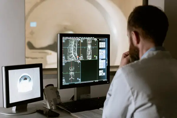
An MRI uses a computer, radio waves, and a magnetic field to capture minute details in a person’s soft tissues. The machine captures nearly every organ image except hard bones. Therefore, a medical professional uses it to view most of your body parts, including your brain and spine.
An MRI detects conditions ranging from tumors to disc hernias or strokes. The machine can also detect traumatic brain injuries, dementia, aneurysms, carotid artery disease, multiple sclerosis, and spinal cord fractures. Read on to understand how powerful these diagnostic machines are, starting with their detail level, types, and how they compare to other medical imaging modalities. Let’s dive in, shall we?
Is an MRI More Detailed Than Other Medical Imaging Modalities?
Apart from MRIs, other medical imaging types include X-rays and CT scans. All these are different medical imaging methods, but they perform an almost similar function of capturing detailed internal images in a person’s body. Healthcare professionals use CT scans and MRIs on different medical grounds. For instance, a CT scan will help diagnose severe spine, head, chest, or abdomen injuries and fractures. Doctors also use them to identify the precise size and location of tumors. X-rays help scan only a few body conditions, including detecting infected tissue.
Unlike MRIs, X-rays and CTs use ionizing radiation in small doses to take and produce internal images. The MRI only uses radio waves and potent magnets to produce the images. Therefore, an individual who undergoes an X-ray or a CT scan is more likely to be exposed to radiation than a patient who underwent an MRI scan. X-rays and CT scans typically take a shorter time to capture diagnostic images than an MRI scan, usually within 5 minutes or less. Conversely, it would take a person up to 30 minutes or more to undergo an MRI scan, depending on what the radiologist is looking for.
MRIs are more efficient in diagnosing ligaments, joints, tendons, and soft tissue problems despite the longer time they take to capture images. They are more resourceful than CT scans and X-rays because they produce clearer 3-Dimensional images. If a doctor needs to see more detailed images of soft tissues and organs, then MRIs are no doubt the best option in this case.
What Is the Level of Detail Involved in MRI Scans?
Your spinal fluid, bone marrow, blood, and soft tissues differ in intensity from black to white when observed under the scan. The intensity varies depending on the machine’s settings and the amount of water and fat in every tissue. A radiologist uses this information to compare the breadth and distribution of the white and black areas, ascertaining whether a tissue is healthy or not.
There are two ways to determine and interpret the results of MRI scans. Medical professionals are best suited in this area, but a little bit of knowledge wouldn’t hurt the regular person. Well, doctors interpret MRI scans according to image view and weight.
Here is a breakdown detailing the two:
1. Image View
Like a Computed Tomography (CT) scan, MRI also produces images in three anatomical aspects. These include the axial, coronal, and sagittal views in a person’s anatomy. A doctor will interpret an axial view from the feet upwards. Therefore, a patient’s right-hand side in the MRI image refers to their left-hand side, and vice versa.
2. Image Weight
Once a doctor has figured out the image view, the next step is to determine the image’s weight. An MRI scanner produces magnetic fields that medical professionals can manipulate to create two image types. The images are referred to as T1 weighted and T2 weighted sequences.
Types of Magnetic Resonance Imaging
There are three types of MRIs, and they include short-bore, standard, and open, as detailed below:
- A standard MRI resembles a long cylinder with a constricted tube in the middle. When patients undergo a scan using this machine, they lie on a movable bed sliding inside the narrow tube. A standard MRI produces pretty impressive images, but patients often complain about feeling claustrophobic while inside the scanner.
- A short-bore MRI scanner is almost similar to the standard one, only that it is most preferred for claustrophobic patients. This machine is half the length of the standard MRI. Therefore, a patient having a head scan will have their feet sticking out one side of the tube. In the case of a back imaging procedure, the head won’t be enclosed in the tube, making the short-bore MRI more bearable during the long scanning process.
- An open MRI machine is ideal for anxious patients or those exceeding the maximum weight requirements other machines have. Unlike the short-bore and standard MRIs shaped inside like a donut hole, the open MRI is designed like a bagel cut into halves. Therefore, it has much more space. The downside to this type is that its images are not as clear as the other MRI scanners.
Magnetic Field Strength
When referring to MRI scanners, it is important to note that the machines have different magnetic field strengths. The unit of measurement that medical practitioners use to identify the field strengths is known as Tesla (T). The higher the tesla scanners, the stronger the magnet in an MRI machine. This strength is usually identified in general and within the machine’s bore. An MRI scanner’s most crucial component is arguably its magnetic field. Most scanners available in the medical field have 3.0 or 1.5 T. Still, some machines also exist with a magnetic flux density below 1.5 T, the most recent one having up to 7.0 T.
A 1.5 T MRI machine has longer sequences that highly improve image quality, while a 3.0 T scanner captures more detail and produces clearer images. Therefore, ordinary scan routines require the standard 1.5 T machine, leaving more complicated imaging like prostrate and spectroscopy to 3.0T machines.
Also, 3.0 T scanners take a shorter time to serve more patients than a 1.5T machine because of their increased signal strength. 7.0T scanners haven’t been influential in a clinical setting yet and are currently confined to research purposes. In 2021, researchers endorsed the first 7T model to operate in clinical settings across Europe and the United States of America.
A 7 Tesla MRI can show small MS plaques and their relationship to cerebral veins for example.
What Is the Strongest MRI That Exists Today?
As of 2018, there were reports of University of Minnesota scientists being the first to perform an MRI scan at 10.5T worldwide. The 10.5 T was the strongest then, having a magnetic field strength 10 times higher than a standard MRI. This strength surpassed even the most advanced magnetic resonance imaging machines present globally in 2018. This scanner held great promise of producing finer details in scans, helping physicians pinpoint the root cause of a problem and prescribe the appropriate medication to patients.
Enter 2021, where another groundbreaking announcement was made: an 11.7T MRI delivering its first-ever images. This machine was touted as the most powerful MRI scanner in the world yet. The machine has a 400-micron resolution in 3 dimensions.
Currently, the world’s most powerful MRI scanner stands at 21.1T. The scanner has a diameter measuring just 10.5 centimeters, too tiny to conduct scans on human beings. The machine is currently at the US National High Magnetic Field Laboratory and only scans small animals. That doesn’t mean it is useless, no. Researchers use the machine to study various patterns, for example, the sodium amount present in rats’ brain tumors. Some of the findings in these studies project that sodium concentration in a tumor reveals its potential to resist chemotherapy treatments.
What Is the Difference Between Detail in Microscopes and MRI?
A microscope is a scientific instrument that individuals use to magnify small objects that the naked eye would find difficult to see. Some microscopes enable scientists to observe more than the surface of an object, helping them see the cellular composition of the object. This includes organelles like the mitochondria, a cell’s shape, and the nucleus.
Microscopes have lenses that enable individuals to see this detail level. Light microscopes influence how light enters the observer’s eye through the convex lens that has both sides bent outward. Therefore, light will reflect off the object and pass through the lens, curving it towards your eye when looking at it. The bending makes the object appear larger than it is. Compound microscopes are the most common microscopes used today. They have interchangeable objective lenses with different magnification powers. The most common powers are 4x, 10x, 40x, and the highest, 100x.
The scanning objective lens (4x) has the lowest magnification power, while the oil immersion objective lens (100x) has the most potent magnification ability. With a 100x objective lens and a 10x eyepiece lens magnification, a compound optical microscope has a 1,000x total magnification. This is the maximum magnification power of the microscope. When comparing an MRI to a microscope, an MRI has greater accuracy levels. This is because samples viewed under a microscope have undergone destructive de-calcification. Adverse de-calcification or sectioning of samples may interfere with their microscopic composition, flawing the optical imaging data. Here are other differences between microscopes and MRI scans:
- MRI scans are used in clinical settings to observe soft tissue anomalies, while microscopes are used in science laboratories to observe specimen samples.
- Microscopes help individuals visualize tiny objects that they don’t normally see, like microorganisms and cells. Conversely, MRI machines help healthcare professionals identify abnormal tissue appearance.
- Individuals use microscopes to observe minute specimens, while MRIs are designed to hold human beings for imaging.
- MRI machines use computers, magnetic fields, and radio waves to capture images, while one can only observe objects under a microscope lens.
Does an MRI Scan Show Everything?
MRI scans can show most body parts, but they can’t show air and hard bone. The two appear black in an MRI image because they don’t signal the machine. Other than hard bones, an MRI shows detailed images of a human’s internal organs like the liver. It also takes the heart, brain, blood vessels, uterus, and spinal cord images.
Below is a video on high Tesla MRI imaging:
Can High Details Be Observed in an MRI Scan of Postmortem Tissues?
MRI scans can conduct postmortem imaging (PMI). PMI is the scanning of autopsy tissues, animals or specimens to identify an individual’s cause of death. This kind of imaging is useful, especially when the deceased’s kin cite cultural reasons that forbid the conventional autopsy. An MRI scan can capture images of postmortem tissues just like live tissue. However, the radiologist has to factor in some changes that may affect the image resolution. The following are stumbling blocks that may hinder a successful postmortem MRI:
- Postmortem changes: These are the natural alterations that occur in a body after death. They include gas accumulation, fluid displacement, blood clotting, and loss of ionic dispersion. When decomposition starts, gas will accumulate inside the deceased’s body, making it extremely difficult to produce high-resolution images showing soft tissue changes.
- The magnitude of the MRI: Conducting an MRI scan on the whole body to find the exact cause of death can be time-consuming and more costly than an autopsy.
- Training and literature: Most of the study material available is based on before-death research findings. Radiologists will compare normal images they saw during the expanse of their career to the MRI image they are currently observing. Therefore, they understand what normal tissues look like, helping them easily spot abnormalities in affected tissues. However, death challenges the norm, making it more difficult to spot conclusive deviations, especially if the radiologist hasn’t specialized in forensic radiology.
- Formalin fixation: Formalin is a substance used to preserve a deceased person’s body. Formalin fixation prevents tissues from undergoing autolysis by changing their microstructure and crosslinking proteins. Certain formalin compositions significantly affect MRI signal tendencies after the fixation.
Click the following link to learn how to improve MRI quality.
Recent Posts
Mastering point cloud to 3d model conversion can feel like translating whispers from another dimension into vivid sculptures. You've got this cloud of data points, a chaotic concert of coordinates...
Let's say you've got a drawing, something you sketched out during a burst of inspiration, and now you're itching to see it leap off the page into three dimensions. Well, that’s exactly what I did...