Wax 3D printing goes back to the 1990s when it was used to create prototypes as well as for the creation of master molds for lost wax casting. Since then, the technology has evolved and expanded. Now you can use wax to create very precise 3d models with intricate geometry in different colors and with different properties.

Let’s look at the technologies involved and steps that need to be taken.
Continue reading “The Complete Guide: How To 3D Print Wax”
How to do 3D Animal Modeling – A Tutorial
Creating a quality 3D animal model includes various steps that need to be taken in a specific order to achieve a realistic and lifelike animal. If you are new to 3D animal modeling or have never tried it before, it may be confusing to figure out what the most important steps are and the order in which they need to be taken.
We’ve compiled all the information you need to create a model. Follow the steps listed below to learn how to do 3D animal modeling:
- Collect Quality References of Your Animal
- Use Basic Shapes to Build the Animal’s Skeleton
- Map Out Your Animal’s Musculature
- Sculpt the Base Mesh Structure of the Body
- Use Retopology to Bring Your 3D Model to Life
- Add Surface Details
How do I Create a 3D Animal Model?
Creating a 3D animal model will take a considerable amount of time, concentration, and commitment. You will most likely need to re-work your model dozens of times before you are satisfied with your final product. However, if you take the time to study your animal and follow the steps listed to create your model, you’ll be well on your way to a high-quality 3D animal model.
Collect Quality References of Your Animal
The better you understand the shape and movement of the animal you’re modeling, the more anatomically accurate your result is going to be. Take the time to collect and study quality references of your animal so that you’re familiar with its anatomy from every angle. Below is a list of different ways you can study your animal, depending on the needs of the project, to get a better idea of how you’re going to represent it in its final 3D version.
- Familiarize Yourself with Your Animal’s Anatomy
- Understand How Your Animal Moves in its Habitat
- Pay Attention to Small Surface Details
- Study Your Animal in Every Stage of Life
Familiarize Yourself with Your Animal’s Anatomy
When it comes to 3D modeling and 3D printing, remember the 80/20 rule: 20% of the work you do before you begin sculpting will determine 80% of the final outcome of your model. Being familiar with the anatomy of the animal you are modeling can be the difference between a mediocre and a lifelike final product. The more accurate you are when creating the inner pieces of your model like the skeleton, the more lifelike the outer pieces will be.
Understand How Your Animal Moves in its Habitat
The way an animal moves plays a major role in its physical presentation. For example, a leopard, which moves very sleekly and stealthily, should be posed differently than an elephant, which moves much more clumsily. This is especially important if you are planning to do any animation but is a great idea to do regardless.
Pay Attention to Small Surface Details
While anatomy and movement patterns are hugely important, don’t forget to pay attention to smaller surface details, such the animal’s fur and skin. Is their fur thick or thin? Does it lay all in one direction, or does it shoot out in many directions? Is their skin soft and smooth, or thick and wrinkly? It can be easy to overlook these small details, but they can be the difference between a good model and a lifelike one.
Study Your Animal in Every Stage of Life
A 3D model of a baby elephant is going to be created very differently than that of an adult elephant. It’s important to study and understand the differences between your animal’s body form and language in every stage of its life. Being familiar with the physical changes your animal undergoes as it ages will allow you to create a much more realistic model of it in whatever stage of life you choose to create it in.
Use Basic Shapes to Build the Animal’s Skeleton
Before you start you will be better off to import drawings or photos of the animal into your 3d program for reference while building the creature. Two planes should be enough such as side view and front view. How you do this will depend on the software you are using so consult with your manual.
Once you’ve collected enough references to start creating your 3D model, you need to need to begin forming the animal’s shape from the inside out. Therefore, the first layer of your 3D model should be the animal’s skeleton. While you don’t need to build out every bone in your animal’s skeleton, you need to have a basic outline of their skeletal system in order to build the rest of the anatomy around it and make it posable or animatable.
Here is a video:
The most effective way to build your animal’s skeleton is to break the major bones and joints down into more basic joints. Once these shapes have created the basic bone structure of the animal, they can be manipulated and resized to the proper dimensions of the animal’s body. Depending on the software used this may be built into the program or a plugin.
Map Out Your Animal’s Musculature
After the basic skeletal system is in place, it’s time to move on to the musculature. This can mean two things: either the actual underlying muscles if you are doing a muscle model for showing whats under the skin such as a muscle anatomy model, or how the muscles affect the shape of the skin.
Here is an approach to building actual muscles:
Some people will actually build a muscle model first and then throw skin on top of it, but this is only practical in programs where it can be done quickly such as in Zbrush, using Zspheres or Zsphere Sketching.
Zspheres:
Zsketch:
Again, depending on whether you will be showing the actual muscle or not will dictate how much detail and realism you will need vs simple shapes. Blocking out the main muscle groups will usually be enough to create a quality surface 3D model.
Sculpt the Base Mesh Structure of the Body
With the bones and musculature in place, it’s time to use them as reference and begin sculpting out the base structure of the animal’s body. This first layer should be built up just enough to cover the animal’s musculature without hiding the outlines of the major muscle groups. Once you’ve established your base mesh, you will later begin to build up detail.
Here is a base mesh modeling example (this one is straight from drawing):
Use Retopology to Bring Your 3D Model to Life
Building up skin on a 3D model can be a tricky and time-consuming process. One of the ways to speed this up is to use “Retopology.” This is the process of re-creating an existing 3D model to give it more fluid geometry. Essentially, retopology acts as a facelift for your original design by correcting imperfections in the mesh and adding texture in areas that need it.
Retopologizing is essential when cleaning up scan data, building a skin on top of quick simple modeling techniques such as Zspheres and Zsketches, and when you need clean geometry for posing your model or animation. Retopology is most often used on models that are used for animation because it reduces the density of the mesh which is essential for movement.
Here is a video:
Add Surface Details
This step of the modeling process lets you add details and personal touches to create the final look of your animal, and make it realistic. See the list below for the different surface details you’ll want to consider for your model and how to create them. When I use the term “surface details” here, it does not only mean traditional polygon or nurbs manipulation.
- Body Composition
- Skin Texture
- Hair/Fur
- Color and Shading
Body Composition
This is where you decide how your animal’s anatomy will show through in their final model. Do you want your animal to look rotund and well-fed, or lean with outlines of certain muscles visible? You may have to do additional sculpting on certain areas to attain the look that you want.
Skin Texture
Depending on the texture of the skin your animal has, you may need to do some additional sculpting. For example, a Rhinoceros’ skin is thick and creased with deep lines and wrinkles. Additional sculpting will be necessary here to achieve the leathery nature of its skin. Keep in mind that sculpting details makes the geometry heavy and the files large. You may want to look at ways to export the details in ways that will decrease the size such as displacement maps or textures.
Hair/Fur
Adding hair or fur can be a time-consuming task, especially if your animal has fur of different lengths or thicknesses. There are multiple tools and plugins to help you with quickening the process of adding fur for animals with very thick or long fur.
Color and Shading
Coloring and shading animal skin and fur can be a tricky task as well, especially for animals with speckled or striped skin/fur. Just like with hair and fur, you can choose whether you want to use tools in your modeling program or plugins.
Here is a video using Blender:
How do I Know When my 3D Animal Model is Successful?
Even if you’ve successfully followed all of the steps to complete your 3D animal model, you may have to re-work your design many times throughout your building process. This is completely normal and can even help you become more familiar with the tools available in your building platform. You may never be completely satisfied with your first attempt. But the more open you are to accepting the ups and downs of building a 3D animal model, the more likely you are to create a successful model that you can be proud of down the road.
Click the following link for a guide on modeling for 3d printing.
Does 3D Rendering Use CPU or GPU Most?
As the 3D animation industry expands in possibility and popularity, a considerable number of professionals are investing in rendering PCs. If you are interested in picking up a rendering PC for yourself or have recently done so, you probably have a few questions regarding its functionality. One frequently asked question inquires into its usage of CPU or GPU.
3D rendering can use either CPU or GPU rendering. However, CPU rendering is the standard, with most current 3D software using a CPU rendering engine. That doesn’t mean GPU rendering isn’t a viable option, but it’s better suited for less complex tasks.
Although CPU rendering is that standard for most 3D rendering software, there are benefits and detriments to using either CPU or GPU rendering. This article will detail the differences between CPU and GPU rendering and explain why CPU rendering is more commonly used over GPU rendering.
Why Does 3D Rendering Use CPU More Than GPU?
Essentially, CPU is the more frequently used 3D rendering engine because it simply delivers greater overall quality than a GPU could at this point. Most renderers use the CPU, however some like Vray use the GPU and speed things up with GPU acceleration.
Although many CPU detractors point to the speed of GPUs, there are several key areas in which GPUs still lag behind CPUs.
| CPU | GPU |
| Handles complex tasks | Struggles with complex tasks |
| Computer holds up to 64GB of RAM | GPU only possesses up to 12GB of memory |
| Very Precise | Moderate Precision |
This information was accumulated from Easy Render.
At this point in time, CPUs are simply a safer bet when compared to GPUs. CPUs are more precise, possess more memory, and can handle more complex tasks than GPUs, which is a vital trait for 3D rendering.
Despite all this, many are making the switch over to GPU. This is largely due to the quick improvements GPUs have made in recent years, their immense processing cores, and their slightly more affordable price.
Although CPU rendering is today’s standard, this mobilization toward GPUs insinuates that that standard is not set in stone.
CPU vs. GPU: A Comprehensive Analysis
3D rendering is fast-changing, so it is worth examining CPU and GPU rendering as potentially viable options. This next section will compare and contrast the performance of CPU and GPU rendering.
What is the CPU?
An appropriate place to compare the two would be to adequately define both CPU and GPU rendering. First, let’s start with the CPU, or central processing unit, rendering.
A CPU is essentially the brain of any computer. It is the core component that stores all of your computer’s memory and runs all of your computer’s programs. Physically, it is a small chip located on a motherboard, containing billions of microscopic transistors that render every function that the CPU performs.
What is the GPU?
A GPU, or graphics processing unit, is a component that was manufactured specifically to power graphics. The component, first manufactured and dubbed a GPU by Nvidia, is commonly used by gamers to improve their PC games’ graphics and rendering speed. However, GPUs have more recently been used for 3D medical imagery and 3D modeling software like AutoCAD.
CPU Speed
A common criticism of CPU rendering is that it is far slower than GPU rendering. This statement is true if you measure each unit’s ability to process multiple tasks simultaneously.
This is due to the number of core processors each unit possesses. CPUs only used to possess one core processor, but now possess anywhere between 1 to 32 core processors. Although this may seem like a decent amount, 24 is a fairly limited number compared to the number of core processors in GPUs.
The limited supply of core processors are plentiful in their strength, making CPUs particularly adept at serial computing, rendering one process at a time. In this regard, CPUs are quick to deliver sequential renders at an extremely high quality.
However, CPUs cannot match the raw speed of GPUs’ plentiful core processors.
GPU Speed
In contrast to the limited number of core processors CPUs possess, GPUs contain a vast number of, slightly less powerful, core processors. Although these core processors are less powerful on their own than the core processors of a CPU, their sheer abundance makes them roughly 3-5 times faster than those of a CPU.
Boxx tested this by pitting CPUs and GPUs against each other to render the same baseline image. The results surpassed the estimates above. The CPU took 19 minutes and 11 seconds to render, while the GPU took only 3 minutes and 4 seconds. This makes this GPU’s performance 6.2 times faster than its CPU counterpart.
CPU Quality
While GPUs render far more quickly than CPUs, the renders’ quality is not always on par with the high quality renders a CPU can achieve. CPUs can deliver consistently high-quality renders, regardless of their complexity, due to their serial rendering structure and the strength of each core processor.
CPUs are able to sync different tasks together with ease, making them far greater at processing more complex 3D scenes. Although this is particularly important for VFX and 3D animation, 3D medical imaging does not possess the same complexity. The resolution and clarity of 3D medical images must be of high quality, but GPUs are less likely to struggle with such tasks.
GPU Quality
As mentioned in the preceding subsection, GPUs, although exceptionally fast at rendering multiple images, can struggle with syncing these renders together. 3D medical imaging relies more on speed over complexity, so this is not necessarily an issue. However, if you are interested in VFX, animation, or graphic design, this can be a detriment to your work.
CPU Cost
Comparing the costs of CPUs and GPUs is tricky because the two often work in unison. However, GPUs are becoming more popular because you can either cut down on the number of CPUs needed to complete the process or entirely cut out CPUs altogether.
This is crucial due to the cost of CPUs when compared to GPUs. CPUs generally cost thousands and are rarely purchased extrinsic to the hardware that contains them. Therefore, you are purchasing the PC along with the CPU. These purchases exceed the minimal requirements for completing the task.
GPU Cost
In contrast, GPUs are commonly purchased extrinsic to hardware, and popular brands like NVIDIA and MSI sell them for hundreds of dollars. Furthermore, you can integrate GPUs into your personal computer, so there is no need to purchase additional hardware. This is why 3D animation has started to embrace GPUs more as a viable 3D rendering option.
Below is a good video on the difference between CPU and GPU rendering:
Final Thoughts
GPUs are making a push to replace CPUs as the primary option for 3D rendering. However, CPUs still deliver more consistent and qualitatively superior renders than GPUs, so if you are working in a more complex 3D rendering field, it is not time to make the switch over just yet.
GPU producing companies like NVIDIA have actually started producing their own CPUs, and CPU oriented companies like Intel have begun working on their GPUs. This perhaps signifies not a wholesale change but the beginnings of a cohesive partnership. Ultimately, the two processing units work best when their powers are combined.
Click the following link to learn how to convert JPEG to DICOM.
3D Animation of Full Human Brain Anatomy and Function
In this comprehensive 3D animation we go over the anatomy of the brain in detail, from the lobes, gyri and sulci of the cortex, to the nuclei of the basal ganglia, white matter, parts of the brainstem, locations of the ventricles, commissures, and other structures. We look at the anatomy as well as physiology, input/output connections, function and importance of each structure. About 60 structures plus their substructures are shown and described in this half hour long animation.
This animation includes views and descriptions of the amygdala, angular gyrus, anterior commissure, basal ganglia, caudate nucleus, central canal of spinal cord, cerebellum, cerebral aqueduct, cerebrum, cerebral cortex, cingulate gyrus, commissure of fornix, corpus callosum, fornix, fourth ventricle, frontal cortex, globus pallidus, gray matter, hippocampus, hypothalamus, inferior colliculus, inferior frontal gyrus, inferior temporal gyrus, internal capsule, interventricular foramina, lateral geniculate, medial geniculate, medulla oblongata, midbrain, middle frontal gyrus, middle temporal gyrus, occipital lobe, orbital gyrus, pineal gland, pituitary gland, pons, postcentral gyrus, posterior commissure, precentral gyrus, prefrontal cortex, putamen, septum, stria terminalis, substantia nigra, superior colliculus, superior parietal lobule, superior temporal gyrus, supramarginal gyrus, thalamus, third ventricle, tuber cinereum, white matter, and others.
Review:
Amygdala
The pair of amygdala deep in the temporal lobes are part of the limbic system. They are almond shaped. The amygdala are made up of the basolateral complex, the medial nucleus, the cortical nucleus, the central nucleus and the intercalated cell clusters. The amygdala is larger in males. They play a role in emotional responses, decision making, and processing of memory. This involves memories associated with emotional events.
Each amygdala has an independent memory system. The right amygdala is concerned with fear conditioning and declarative memory as well as in associating time and place with emotions. The amygdala are involved in memory consolidation. It is the main structure involved in the fight or flight response.
Projections of the amygdala include the locus coeruleus, the hypothalamus, the nucleus accumbens, and the thalamus. There is data showing that the left amygdala has a role in the reward system. It has also been linked to obsessive and compulsive bevavior, anxiety disorders, and PTSD. The left amygdala develops first, while the right amygdala grows for a longer span of time.
Angular gyrus
The angular gyrus is in Brodmann area 39. It is posterior to the supramarginal gyrus in the parietal lobe, and close to the superior edge of the temporal lobe. It is involved in complex language functions such as reading, writing and their interpretation. It sends visual information to Wernicke’s area. Its other functions involve attention, memory retrieval, and spatial cognition including distinguishing between left and right.
Anterior commissure
The anterior commissure is a white matter tract much smaller than the corpus callosum. It connects the two temporal lobes of the cerebrum in front of the fornix. It is important in pain sensation and olfaction as it includes fibers from the neospinothalamic tract and olfactory tracts.
Basal ganglia
The basal ganglia is a group of subcortical nuclei on top of the midbrain. They include the dorsal striatum (caudate nucleus and putamen), the ventral striatum (nucleus accumbens and olfactory tubercle), ventral pallidum, the globus pallidus, substantia nigra and subthalamic nucleus.
They connect with the thalamus, brainstem, cortex and others.
Caudate nucleus
The caudate nucleus along with the putamen form the dorsal striatum, which is divided by the internal capsule. In each hemisphere, it forms a thick anterior part called the head which tapers to a body and narrow tail, giving it a C shape. The tail curves toward the front. It is located on both sides of the thalamus.
It functions in associative and procedural learning, inhibitory control of action, and movement. There is a link between the caudate and sleep patterns. Movement functions include posture as well as speed of directed movements.
Neuronal connections from the caudate reach the substantia nigra and globus pallidus and it is innervated by neurons from the substantia nigra. The caudate has projections to the hippocampus and receives projections from the amygdala. There are data showing effects on emotion, hyperactivity, and drive.
Central canal of spinal cord
The central canal runs through the spinal cord. It is filled with cerebrospinal fluid and it transports nutrients to the spinal cord. In the upper regions of the spinal cord it can be found in the anterior portion, later becoming more central and ultimately posterior. The widest part of the central canal is the terminal or fifth ventricle.
cerebellum
The cerebellum is located in the posterior underneath the cerebral hemispheres. The pons and medulla are anterior to it. It is part of the metencephalon. The actual name means “little brain”. Its made up of a gray matter cortex, deeper white matter with myelinated fibers, deep gray matter cerebellar nuclei inside the white matter, and a fluid filled ventricle.
The cerebellum is made up of two hemispheres and a protruding midline vermis (worm in Latin). The white matter is called the arbor vitae or tree of life due to its appearance when sectioned. The cortex is made up of a continuous layer of tissue folded repeatedly although it appears as parallel grooves on the surface. The gyri of the cerebellum are called folia.
The lateral cerebellum is called the cerebrocerebellum and the medial cerebellum is called the spinocerebellum. The cerebellum is divided into three lobes- anterior above the primary fissure, posterior below the primary fissure, and flocculonodular below the posterior fissure.
The cerebellum is associated with movement, including motor learning, timing, coordination and precision as well as language and attention. The cerebellum receives input from sensory systems of the spinal cord. Neurons most commonly found in the cerebellum include Purkinje and granule cells. There are 3 important axon types in the cerebellum, mossy fibers, climbing fibers and parallel fibers.
Cerebellar peduncles
Three pairs of cerebellar peduncles connect the cerebellum to the nervous system.
Superior cerebellar peduncle-connects to the cerebral cortex
Middle cerebellar peduncle-connects to pons
Inferior cerebellar peduncle-output to reticular formation and vestibular nuclei
cerebral aqueduct
The cerebral aqueduct is found ventral to the cerebellum and dorsal to the pons. Also called the Sylvian aqueduct, it contains CSF and connects the third and fourth ventricle. The gray matter around the aqueduct is called the periaqueductal gray.
cerebrum
The cerebrum or telencephalon is the largest part of the brain. It contains not only the two cerebral hemispheres made up of an outer cortex of gray matter and inner white matter but also subcortical structures such as the basal ganglia, hippocampus and olfactory bulb. The cerebrum is involved in voluntary motor actions, sensory perception, memory and thoughts. The cerebrum develops from the prosencephalon.
Cerebral cortex
The cerebral cortex is the outer gray matter layer of the cerebrum and is made up of two hemispheres. The 2 cerebral hemispheres consist of depressions or sulci and elevations or gyri. It is partially separated by a deep longitudinal fissure. The medial longitudinal fissure separates it into the two hemispheres.
The cortex can be divided into the frontal, parietal, occipital and temporal lobe. The cortex includes primary sensory areas which receive sensory information and association areas. The frontal lobe includes Broca’s area responsible for production of language and the temporal lobe includes Wernicke’s area responsible for speech comprehension.
choroid plexus
The choroid plexus is a plexus of cells that produce and secrete cerebrospinal fluid. It also acts as a blood/CSF barrier. It is made up of a core of capillaries and connective tissue surrounded by cuboidal epithelial cells. Every one of the four ventricles contains a choroid plexus.
cingulate gyrus
The cingulate cortex includes the whole cingulate gyrus and can be found medially in the cerebral hemispheres above the corpus callosum. It is part of the limbic system. It functions both in respiratory control, executive function, as well as learning, memory and emotions.
commissure of fornix
The commissure of fornix is formed by fibers left and right fornix bundles.
corpus callosum
The corpus callosum, which translates into “tough Body”, is a thick nerve tract that connects the two cerebral hemispheres. It can be found below the cerebral cortex crossing the midline. The corpus callosum is the largest white matter body with several hundred million axons. It can be divided into the rostrum, the genu, the body and the splenium. The corpus callosum allows for communication between the two hemispheres of the cerebrum.
fornix
The fornix is a part of the limbic system and is an output of the hippocampus. It starts on each side in the hippocampus as fimbria hippocampi and continue as the crura or posterior pillars. At the commissure of fornix the fibers meet forming the body. Around the anterior commissure the body divides again into the anterior pillars or columns which continue on to the mamillary bodies. The fornix is important in establishing episodic memories and fornix damage leads to problems with spatial memory.
fourth ventricle
The fourth ventricle carries cerebrospinal fluid and can be found extending from the aqueduct of Sylvius to the obex in the caudal medulla. It is at the level of the pons. The cerebellar peduncles form the sides of the fourth ventricle. The rhomboid fossa is the floor of this ventricle. The fourth ventricle has a diamond shape in cross section. The CSF flows into the fourth ventricle through the cerebral aqueduct.
frontal cortex
The frontal cortex as the name suggests can be found at the anterior of each cerebral hemisphere. It is separated from the parietal lobe by the central sulcus and from the temporal lobe by the lateral sulcus. The premotor as well the primary motor cortex are found in the frontal cortex.
globus pallidus
The globus pallidus which is part of the basal ganglia, translates into “pale globe”. There are two parts: The pars externa and pars interna. It projects mainly to the substantia nigra and thalamus and functions in regulation of voluntary movement, through inhibitory action, whereas the cerebellum provides excitatory action. It receives inputs from the caudate and putamen.
gray matter
Gray matter which consists of cell bodies includes the basal ganglia including the nucleus accumbens, caudate nucleus, putamen, and substantia nigra as well as the surface of the cerebrum and cerebellum, thalamus, hypothalamus and brainstem nuclei. It is gray colored due to the color of neuron cell bodies as opposed to white matter which appears white due to the color of myelin.
habenula
The habenula is a cell mass on the side of the third ventricle located above the thalamus. It is called the crossroad between the basal ganglia and the limbic system.
hippocampus
The hippocampus, or seahorse in latin, is part of the limbic system and can be found as a pair, one on each side, by the floor of the lateral ventricle. The parahippocampal gyrus conceals the hippocampus. It is made up of the hippocampus itself and the dentate gyrus, which is surrounded by hippocampal gray matter.
The name cornu ammonis or ram’s horn is given to it because of its shape as shown in cross-section. The abbreviation CA given to its parts including CA1, CA2, CA3 and CA4 come from this name.
The hippocampus takes part in storing of explicit memory, such as information about objects, places and people. This is done through long term potentiation. Hippocampal damage is seen in dementia such as Alzheimers disease. It is also affected in schizophrenia and PTSD.
The three major pathways in the hippocampus include the mossy fiber pathway, the schaffer collateral pathway and the perforant fiber pathway. Input comes from the entorhinal cortex. Outputs include the entorhinal cortex and the prefrontal cortex. It is modulated by dopamine, serotonin and norepinephrine. The hippocampus is one of the few brain regions where new nerve cells are born.
hypothalamus
The hypothalamus is part of the limbic system and is found under the thalamus. The name hypothalamus means “under chamber”. The hypothalamus links the nervous system and the endocrine system via the pituitary. Therefore it has a neuroendocrine function. It works to control sleep, hunger, body temperature and activities of the autonomic nervous system. The hypothalamus synthesizes and secretes hormones which control the pituitary gland. Its connections include the reticular formation, brainstem, amygdala and septum. Other functions of the hypothalamus include controlling defensive behaviors.
inferior colliculus
The inferior colliculus is located over the trochlear nerve and below the superior colliculus. It is a large main nucleus of the auditory pathway in the midbrain where auditory pathways converge. It also functions in spatial localization through hearing as well as a integration station and switchboard. Its inputs include the auditory cortex and brainstem nuclei. The name inferior colliculus means lower hill. Inputs include several brainstem nuclei.
inferior frontal gyrus
The inferior frontal gyrus, a part of the prefrontal cortex, is the location of Broca’s area, responsible for speech creation and language processing.
Inferior temporal gyrus
The inferior temporal gyrus is the lowest of the gyri of the temporal lobe. It is separated from the middle temporal gyrus by the inferior temporal sulcus. The occipital temporal sulcus separates it from the fusiform gyrus. It is involved in visual stimulus processing on the level of object recognition based on form and color.
internal capsule
The V shaped internal capsule is a white matter structure that separates the caudate nucleus and putamen and also the caudate and thalamus from putamen and globus pallidus. It contains ascending and descending tracts connecting the cortex. The bend in the V of the internal capsule is called the Genu. It has an anterior limb and a posterior limb. Much of the internal capsule is the corticospinal tract which carries information from the primary motor cortex to motor neurons in the spinal cord.
interventricular foramina
The interventricular foramina, also called the foramina of Monro, are channels which connect the right and left lateral ventricles to the third ventricle. Cerebrospinal fluid flows through the interventricular foramina into the third ventricle. They contain a choroid plexus in their walls which produces CSF.
lateral geniculate
The lateral geniculate is a small thalamic nucleus located at the end of the optic tract. It is a visual pathway relay center. Information from the eye and optic tract goes through the lateral geniculate on its way to the primary visual cortex. The lateral geniculate has 6 layers of neurons.
lateral ventricle
There are two C shaped cerebrospinal fluid filled lateral ventricles. They stretch from the inferior horn of the temporal lobe all the way down to the third ventricle. They can be divided into three horns and a body. The frontal horn can be found in the frontal lobe of the brain while the occipital horn enters the occipital lobe. The lateral ventricles are the largest of all the ventricles.
medial geniculate
The medial geniculate body is made up of several nuclei. Its part of the auditory system. The MGB is a relay between the inferior colliculus and the auditory cortex.
medulla oblongata
The medulla oblongata is part of the brainstem. Medullary pyramids contain the corticospinal and corticobulbar tracts. The swellings or olives are due to the inferior olivary nuclei. The medulla connects higher brain levels to the spinal cord. The medulla functions in involuntary actions. Its autonomic control includes heart rate, respiration, sleep and heart rate.
midbrain
The midbrain is the anterior part of the brainstem. The dorsal side of the midbrain is the tectum, which means roof. The tectum has 4 colliculi or bumps on its surface. The tegmentum is the floor and is ventral to the cerebral aqueduct. Its involved in homeostasis. Superior colliculi process visual information and inferior colliculi process auditory information. It functions in functions in alertness, sleep, temperature regulation, hearing and vision.
middle frontal gyrus
The middle frontal gyrus is found between the superior and inferior frontal sulci. It has a role in the reorienting of attention.
middle temporal gyrus
The middle temporal gyrus is located in the middle of the temporal lobe. It has been linked to accessing word meaning while reading, figuring out distance, and recognition of known faces.
occipital lobe
The occipital lobe of the cerebral cortex is named after the occipital bone and is the smallest lobe of the cortex. It is located above the temporal and below the parietal lobe. The occipital lobe includes the visual cortex, mainly Brodmann area 17 or V1. It also includes V2 or the ventral stream, which is a secondary visual cortex. Underneath the occipital lobe is the tentorium cerebelli which divides the cerebellum from the cerebrum. The lateral occipital sulcus separates occipital gyri.
orbital gyrus
The orbital gyrus can be found on the inferior surface of the frontal lobe. This area of the front lobe rests on the orbital plate of the frontal bone. There are 4 orbital gyri- the posterior, lateral, medial and anterior.
optic chiasm
The name optic chiasm comes from the greek for crossing. It is X shaped. It can be found under the hypothalamus and is where the optic nerves cross. Fibers from the medial part of the retina cross over here but fibers from the lateral half of the retina stay ipsilateral. It is located in the chiasmatic cistern and is encircled by the circle of willis.
optic tract
The optic tract is found in a pair and is an extension of the optic nerve. It conveys information to the contralateral half of the visual field. Axons of the right and left optic tracts synapse at the lateral geniculate nucleus.
pineal gland
The pineal is a small endocrine gland which is not paired. The name comes from the shape of the gland, which looks like a small pine cone. It produces melatonin, a serotonin derived hormone, that modulates sleep cycles. The pineal can be found where two of the halves of the thalamus meet behind the third ventricle and is part of the epithalamus.
pituitary gland
The pituitary is an endocrine gland located at the bottom of the hypothalamus. Hormones secreted here control blood pressure, temperature regulation, sex organ function, thyroid function, pain relief and water regulation. The posterior pituitary is connected to the hypothalamus by the pituitary stalk. The pituitary has an anterior, intermediate and posterior lobe.
pons
The word pons comes from the Latin for bridge. It is in the brainstem between the midbrain and the medulla. Within it are tracts that carry sensory signals up and other signals down. In the rear it is made up of two pairs of cerebellar peduncles. The middle cerebellar peduncle connects the pons to the cerebellum.
It is located in front of the cerebellum. It has two major divisions- the ventral pons and the tegmentum. The pons contains several cranial nerve nuclei including the vestibulocochlear nucleus, facial nerve nucleus, nucleus abducens and the motor nucleus of the trigeminal nerve. It is part of various autonomic functions such as arousal, sleep regulation, equilibrium, and muscle tone.
postcentral gyrus
The postcentral gurys is located at the anterior parietal lobe. It includes Brodmann areas 1, 2, 3. It is where the primary somatosensory cortex can be found. It is the location of neurons that integrate sensory information from distinct parts of the body. In front of it is the central sulcus. In the back of it is the postcentral sulcus.
posterior commissure
The posterior or epithalamic commissure is a white matter tract which connects the two hemispheres of the cerebrum. It crosses the midline dorsally of the cerebral aqueduct. It forms one of the stalks that attach the pineal gland to the wall of the third ventricle. The posterior commissure connects language processing centers of the two hemispheres.
precentral gyrus
The precental gurys is Brodmann area 4. It is located at the rear of the frontal lobe just before the central sulcus. It has a diagonal orientation and is continuous with the postcentral gyrus. Behind it is the postcentral gyrus. This is the location of the primary motor cortex involved in execution of voluntary motor movements and skeletal muscles.
prefrontal cortex
The prefrontal cortex can be found in the anterior frontal lobe. It is where executive function is carried out and high level filtering.
meaning acceptable behavior, predicting outcomes, determination of good and bad, working toward a goal. It receives connections from brainstem arousal systems.
putamen
The putamen is a round structure that is a part of the basal ganglia. Together with the caudate nucleus it forms the dorsal striatum. To the medial of it lies the globus pallidus. It functions in regulation of movement and is connected to the globus pallidus and substantia nigra. The putamen is affected in Parkinsons disease where involuntary muscle movements occur. Together with the globus pallidus it is known as the lentiform nucleus as they appear like a lens like shape.
septum
The septum pellucidum is a membrane between the two cerebral hemispheres. It separates the lateral ventricles and encloses the fifth ventricle. It stretches from the corpus callosum to the fornix.
stria medullaris of hypothalamus
It is part of the epithalamus and is a horizontal ridge on the medial surface of the thalamus.
stria terminalis
The stria terminalis or the terminal stria is a bundle of fibers from the amygdala to the septal nuclei, hypothalamus and thalamus. It runs across the lateral wall of the intra ventricular surface. It marks the separation between the thalamus and caudate nucleus.
Substantia nigra
It is a long nucleus in the midbrain, however its functionally a part of the basal ganglia. The substantia nigra plays roles in regulation of movement and muscle tone. There are two parts in the substantia nigra: the substantia nigra pars compacta and the substantia nigra pars reticulata.
The substantia nigra pars reticulata is more anterior and contains GABAergic neurons. The Substantia nigra pars compacta is very populated by dopaminergic neurons. In Parkisons disease, the pars compacta degenerates. Both parts receive input from the caudate and putamen whereas the pars reticulata sends information outside the basal ganglia.
superior colliculus
The superior colliculus can be found on top of the midbrain and is a paired structure. It is a multi sensory structure. Some of its functions include directing eye movements, head turns, and shifts in attention.
superior parietal lobule
The superior parietal lobule includes Brodmann area 5 and 7. Its functions include spatial orientation. It receives visual input and sensory input.
superior temporal gyrus
The superior temporal gyrus is located in the temporal lobe below the lateral sulcus. It includes brodmann area 41 and 42 and Wernickes area. It is the location of the auditory cortex and processing of sounds.
supramarginal gyrus
It is located anterior to the angular gyrus. It is part of the somatosensory association cortex. Its involved in the interpretation of tactile sensory data.
Thalamus
Its a gray matter structure in the forebrain. It is found medially. The thalamus is a hub for information relay. It relays sensory signals, regulates alertness, arousal, sleep and consciousness. Every sensory system utilizes the thalamus except for the olfactory system. Its connected to the cortex by many thalamocortical radiations.
third ventricle
It is filled with CSF and found between the two halves of the thalamus. It is narrow. The epithalamus is behind it.
tuber cinereum
The tuber cinereum, sometimes called the pituitary stalk is a hollow eminence of gray matter. It contains fibers going from the hypothalamus to the pituitary gland. It is part of the hypothalamus.
white matter
White matter is found deep in the brain including the cerebellum and superficially in the spinal cord. White matter is made up of axon bundles that connect gray matter areas. Within the white matter one can find gray matter nuclei like brainstem nuclei and the basal ganglia. The name comes from the color of fatty myelinated axons.
Click here for a 3d animation of human kidney structure and function.
How to Email DICOM Images in 3 Steps
Medical imaging files carry a lot of information in them, which usually makes the DICOM file size too large to attach to an email.

The faster and easier they can be shared, the better though, and there are still ways to share DICOM images electronically.
Continue reading “How to Email DICOM Images in 3 Steps”
Does Redshift Work with Blender?
Both Redshift and Blender are currently rising within the 3D field. With Blender offering unmatched modeling software in an open-source format, they are the perfect platform to make use of Redshift’s GPU-accelerated rendering software.
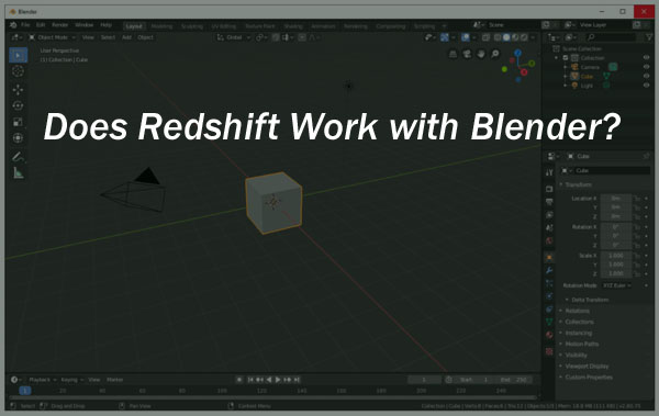
While it has not seen a full commercial release as of November 2020, Redshift support for Blender was in open beta for the public to test. Maxxon’s 3.0.33 update was the first to offer the Blender support beta test. Developers were working on an official plugin that was still in the middle of its development.
While it is not yet feature-complete, this release shows that the developers at Maxxon were working on it. Despite its early testing phase, moving your creations and trying out the new renderer can offer some incredible speeds. If you’re interested in how Redshift will work with Blender, keep reading.
