
The confocal microscope is an amazing and useful piece of equipment, allowing one to look into tissues with greater clarity and without the limitations of other microscopes. Not only can it be used to exactly pinpoint your stained objects of interest, but the 3D stacks it is capable of producing can be turned into virtual 3D worlds and animations you can fly through as if you were inside the living tissue.

However, even the most knowledgeable science professionals sometimes miss out on the essential details. You could own a piece of certain laboratory equipment and remain unsure of its full potential. In this article I will discuss the maximum magnification of a confocal microscope and related items.
In general, the maximum magnification of a confocal microscope is 1000x, assuming the use of a combination of a 100x objective and a 10x ocular. It is dependent on the limitations of the mounting type used, thickness of tissue, and optics of the system.
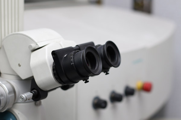
Let’s discuss this in detail. Confocal microscopes much like other microscopes work using interchangeable objectives, which in combination with the eye oculars of a certain magnification, provide the final magnification of the object. This is because you are looking through both at the same time and both must be added up. Oculars are not changed and usually have a 10x magnification.
The objectives themselves can be anywhere from 1x to 100x, depending on the needs of the observer and on the magnification limit of the tissue and other factors such as thickness of slide. This gives a total magnification range of 10x to 1000x. Objectives with higher magnification are of a special type, and require immersion in oil or water, due to the fact that light does not travel as well through air.
As a comparison, total maximum magnifications are 1,000,000x for electron microscopes and about 1500x for a light microscope. Scanning electron microscopes have smaller maximum magnifications than transmission electron microscopes. Each type of microscope, however, has its limitations, whether its in the optics or the way the tissues are prepared or mounted.
Factors affecting magnification
As with any microscopy image acquisition, garbage in means garbage out. You will not be able to magnify a poorly prepared sample to a large degree. Proper preparation includes correct tissue processing, fixation and labeling. Your protocols must be optimized and fit the type of tissue you want to examine.
The specimen mounting is somewhat dependent on the tissue of interest- is it a fixed mount, hardened or liquid? For 3D stacks, you may want to keep the specimen in liquid media as media which harden make the sample flatter.
The thicker the tissue you use, the more difficult it will be for light to pass through, and in addition the less powerful an objective you will be able to use, as larger magnification objectives have a shorter working distance. These are two reasons why thicker tissues limit magnification options.
Maximum resolution of a confocal microscope
The resolution of a microscope is related to magnification. Resolution of a microscope refers to the minimum separation between two points that can be seen by the operator. For a confocal microscope, the maximum resolution is around 500 nanometers axially and 150 nanometers laterally.
Resolution in the case of a confocal microscope is dependent not only on the optical components but also on the wavelength of incident and detected light. In fact, shorter wavelengths give higher resolution.
For comparison, the maximum resolution for an electron microscope is 0.2 nanometers, while for a light microscope it is about 200 nanometers.
How does a confocal microscope work
Unlike stereo and compound microscopes which use regular light for image formation, the confocal microscope utilizes a laser beam to scan samples that have been dyed. These samples are prepared on slides and inserted; then, aided by a dichromatic mirror, the device produces a magnified image on a computer screen.
A confocal microscope, known in some circles as a confocal laser-scanning microscope, is an epifluorescent microscope. Confocal microscopy harmonizes laser source for fluorescence excitation instead of the traditional metal halide lamp or LED used in a wide-field epifluorescent microscope.
The fundamental operation of a confocal laser-scanning microscope is identical to that of a wide-field epifluorescent microscope. The excitation laser beam is irradiated to the fluorescent sample, which gives off fluorescence transmitted through the microscope’s objective lens. The barrier filters subsequently separate the fluorescence from the scattered and stray excitation photons before reaching the pinhole. The pinhole then eliminates emitted fluorescence detected from optical planes in the specimen other than the focal plane. Finally, the PMT detectors relay the detected photons as an electric signal to the connected computer terminal.
However, as the laser source used to excite the specimen essentially emits just a single spot (think of your laser pointer, which emits a single bright spot of light), two galvo mirrors in the X- and Y- axis rapidly scan the laser spot across the specimen. These mirrors work to obtain a single plane of illumination so that any fluorophores contained within the excitation plane of the fluorescent-labeled specimen would emit fluorescence. The computer terminal, which is connected to the PMTs in the detector, then processes the received electrical output into a final 2-dimensional page which the user can observe through the screen or monitor.
What can you see with a confocal microscope
Biologists use a confocal microscope to detect numerous microorganisms. It has been used earlier to observe hidden microbes on surfaces, in water, and other habitats. Confocal microscopy has been used in the study of many diseased and healthy tissues, from the brain to the liver.

Confocal microscopy plays a crucial role in detecting corneal infection-causing microbes. Organisms like Acanthamoeba (animal-like protists) are tough to bring up on ocular surfaces since their impression can imitate bacterial keratitis. With confocal microscopy, the acanthamoeba appears as a circular or ovoid highly-reflective substructure. Confocal microscopy is also used for disease diagnosis.
Why is a confocal microscope unique?
The confocal microscope is known for its efficiency and its utmost importance in the field of research. With the utility of confocal microscopy, researchers are afforded an edge and a massive advantage in performing efficient and more accurate research work. This is because confocal microscopy presents the ability to collect serial optical sections from thick specimens, thereby creating detailed images of sample tissues or objects.
Fluorescent tagging in confocal microscopy is especially relevant when rendering images as it allows very distinct segregation of structures and the ease of perception of their different locations. Confocal microscopes can also produce three-dimensional images and two-dimensional visualizations most microscopes provide.
This illumination, therefore, includes a rapid, non-invasive technique that presents an early diagnosis and management coupled with high-resolution images compared to CT scan, MRI, and USG for dermatological use. Confocal microscopy affords the research firsthand and detailed images of the innermost composition and structure of the cells and tissues of the human body. This confocal microscope is usually employed in the dermatology and skincare industry for both research and diagnostic purposes.
Where is a confocal microscope used?
Confocal microscopy is a valuable image tool that is beneficial to study the biomolecules applied to the skin. It is utilized by pharmaceutical researchers to design and optimize skin delivery systems, as well as to diagnose skin diseases. The confocal microscope is adopted to characterize pharmaceutical systems, including tablets, film coatings, and colloidal systems.
Confocal imaging is utilized in the study and the investigation of the structure and time evolution of transient gels, which are formed in the mixtures of colloid polymers. Visualizations of the structures within thicker objects can conveniently be achieved using these systems. Essentially, every medical establishment with high stakes and concentration on both research and scan analysis utilizes confocal microscopy in achieving results.
This laser scanning microscope is commonly employed within pharmaceutical companies that deal majorly in skincare products and skin health. This is because of the standard and high definition imagery it boasts of due to its fluorescent attribute. Semi-conductor industries, as well as life and material laboratories, also make use of these microscopes. Its utility is also evident in studying and evaluating live cells, thus making it essential equipment of every biologically inclined research facility or laboratory.
Who can use a confocal microscope?
Biology, as it is well known, is the study of life, and while on this study, viewing the objects being studied goes a long way. A confocal microscope is a vital tool for biologists as it allows them to study the composition and structures of live cells firsthand. Researchers also involved in material science can utilize this microscope for a better understanding and assimilation of research methodology and processes.
Persons involved in the semi-conductor industries can also use the confocal microscope for efficient and effective delivery and result actualization. As common and as simple as it sounds, this laser scanning microscope, however, requires a form and level of professionalism and experience in its handling. It can be somewhat confusing to one not too familiar with other types of microscopes.
High-resolution imaging helps microbiologists obtain a better understanding of the morphology of microbial populations, biofilm formation, and also communication among bacteria. For zoologists, situ experiments are viewed in three-dimensional imagery, offering them more observatory experience with the utility of a confocal microscope.
When can a confocal microscope be used?
In any case of a need to examine the detailed structure of specific objects within the cells of both plants and humans, the confocal microscope can be utilized. Research into the functions and characterization of the inner workings of animal cells can be conducted with the help of these microscopes.
Also, in the event of meticulous observation and study of the makeup and structures that exist within cells and tissues, the confocal microscope can be utilized effectively as it offers imagery that presents details in their purest form. This way, little sights which would ordinarily escape the human lens and the lenses of other microscopes are caught and depicted as they truly exist.
Limitations of the confocal microscope
As essential as this image tool is, there are specific problems with which it is associated. Fundamental, also, is the fact that confocal microscopes operate with a mechanism that requires a compromise between resolution, scan time, and photo destruction of the specimen. This implies that the higher the resolution, the more time it requires to scan and the longer the exposure time. In many instances, the resolution enhancement does not increase the essential biological information derived from the specimen. There is also the problem of phototoxicity and photobleaching and a high provision for the misuse of the instrument in the field of research.
You can learn more about confocal microscopy by watching this video by Global Bioimaging:
In summary
The confocal microscope is a huge blessing to both medical establishments and research laboratories. It has contributed immensely towards the effective actualization of results and successful conduct of experimental procedures.
It is, however, not limited to just the medical or biological areas of science. The importance of confocal microscopy cuts across various aspects of life and into other areas of science and technology, most of which are foundational aspects of our world today.
I hope this article on confocal microscopy was useful.
Click the following link to learn the differences between Amira and Imaris.
Recent Posts
Mastering point cloud to 3d model conversion can feel like translating whispers from another dimension into vivid sculptures. You've got this cloud of data points, a chaotic concert of coordinates...
Let's say you've got a drawing, something you sketched out during a burst of inspiration, and now you're itching to see it leap off the page into three dimensions. Well, that’s exactly what I did...