
3D reconstruction software is truly a game-changer for scientific visualization. As technology has improved over the past decade, medical professionals have seen huge advancements in the data sets provided by CT-Scans, MRIs, and medical ultrasounds as a result of this software.
Let’s discuss this topic in detail.
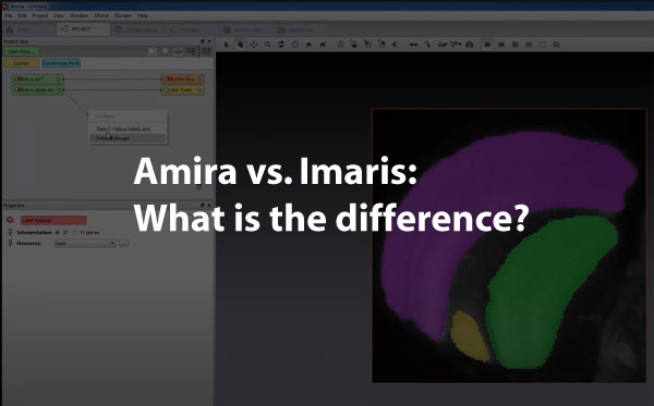
Two well-known names in biological 3D visualization software are Amira and Imaris. While very similar, Amira is superior when working with large data sets, and its strength is using source images from medical scans such as MRI or CT. Imaris offers great specialized options for different fields of study but still provides robust performance in 3D/4D imaging. Imaris excels when using microscopy data such as confocal.
With many 3D Visualization software programs available today, it can be overwhelming when trying to choose the right product to fit the specific needs of an organization, department, or researcher. Amira and Imaris are both solid choices for a variety of applications, as seen below.
An Overview of Amira
Amira 3D visualization products were launched in October of 1999 by Thermo Fisher Scientific. The Amira product line includes 3D – 5D visualization tools and software for several industries, including medicine.
The Amira Life & Biomedical Sciences
This Amira product is the visualization tool used for auto-segmentation, object separation, automatic labeling. Amira does not offer package options for different areas of expertise. This all-in-one product is designed for a range of medical professionals, including
- Physicians
- Researchers
- Cell biologists
- Neuroscience professionals
Features and Capabilities of Amira’s Life & Biomedical Science Product
| Importing and Processing Image Data |
● Able to handle large data sets ● Offers image enhancement, comprehensive filtering ● Scales, calibrates, and converts data
|
| Visualization and Tracking |
● Interactive, multichannel visualization ● Molecular visualization ● Single-cell tracking
|
| Segmentation |
● Auto-segmentation, object separation ● Automatic labeling ● Automatic tracing of individual fibers and filaments ● 3d surface reproduction
|
| Analyzation/Quantification
|
● Convenient built-in measurements
● User-defined measurements can be created ● Results viewer with a spreadsheet and charting tool
|
| Presentation | ● Animation and video available
● Export images, 3d models, spreadsheets ● Single and tiled screen display |
Feedback from Amira Users
- Compatibility. Amira, like Imaris, is compatible with Python and MatLab.
- Ease of use. Users of Amira often cite ease of use as the primary benefit of the tool. With an extremely user-friendly interface, Amira is a great choice for researchers and professionals who may not be particularly computer savvy.
- Free trial. Amira offers an open-source, free trial period.
An Overview of Imaris
Introduced in 1993, Imaris has been an industry leader for many years, and its products have evolved over time to fit the needs of a variety of applications. They offer several different packages to meet the needs of their different clientele.
| Imaris Start | Imaris for Tracking | Imaris for Cell Biologists | Imaris for Neuroscientists | |
| Provides 3D/4D imaging
|
Yes | Yes | Yes | Yes |
| Provides detailed object measurements, reporting, and analysis
|
Yes
|
Yes | Yes | Yes |
| Visualizes and quantifies colocalized regions
|
Yes | No | Yes | Yes |
| Plots 1D-4D, allows for comparisons with statistical tests
|
Yes | No | Yes | Yes |
| Tracks motion in 2D/3D
|
No | Yes | Yes | Yes |
| Allows for tracing filaments, neurons, vessels
|
No | No | No | Yes |
| Allows for segmentation/analyzation of cells & cell structures
|
No | No | Yes | No |
| Customizable analysis with Matlab, Python, Java
|
No | No | Yes | Yes |
| Provides Alignment and stitching of tiles | Optional feature | Optional feature | Optional feature | Optional feature |
Feedback from Imaris Users
- Overall performance and flexibility. These strengths of the program often cited, especially in data analysis.
- Reporting and data analysis functions are fully customizable and compatible with users in Python, as well as Matlab.
- Free trial. New users can get a free trial of Imaris, with all available features to determine what product to choose and which features are useful for their specific application.
Note: Imaris also offers a product that features all of the capabilities listed above called Imaris Single Full for professionals who want all of the capabilities that exist for each product line.
Similarities between Amira and Imaris
The Amira Life & Biomedical 3D Visualization platform is very similar to the Imaris Single Full product.
Both programs offer an open-source, free trial period, where users can download the program and try out the features of the program(s) and determine which is the best fit.
Both Amira and Imaris are both highly rated programs that can provide physicians, researchers, and other medical professionals with a wealth of information and process large data sets, but each program has its individual strengths.
Why Choose Amira?
Amira provides users with a wide range of capabilities, and when compared to similar products, is superior when dealing with very large data sets.
The larger the data set, the more complex the end result, and across the board, from professionals in different Life/Science fields of study, Amira gets high marks in this area.
Pros and Cons of Amira
- Higher cost. Amira is definitely on the higher end in terms of pricing.
- Inflexibility. Without the flexibility of Imaris product offerings, some Amira end users may end up paying for a ton of features that they may not want or need.
- Good for large, varied organizations. In research roles, hospitals, educational institutions, or other large organizations, Amira provides users with a ton of options and capabilities.
- Ease of use. Amira is based on an interconnected node system where each new function graphically appears as a node, similar to Maya’s hypergraph. This way you always have a visual graph of your progress and can go back and delete or modify nodes.
Why Choose Imaris?
With several program options, users can select a product that gives them exactly what they need, without a lot of extra, unnecessary capabilities.
The specialization of Imaris options helps streamline staff training and keeps costs much lower, which can be a huge factor in some industries, particularly education.
Where Amira Excels
- 3D/4D visualization and tracking. Imaris gets top marks for their 3D/4D visualization, especially with tracking objects and single cells.
- High-quality graphics and visualization. The Imaris Cell Biologist program gets high marks from end-users due to the excellent tracking and overall visualization, as the program provides high-quality graphics and animations.
- Excellent for small subjects. For working with single cells, neurons, filaments, and other small subjects, Imaris is a wise choice.
Why the Right 3D Visualization Software Is Crucial
3D Visualization tools have helped lead advances in research, surgical procedures, radiology, and health education. With access to more information about the human body, from single-cell components to full-body systems and individual organs, 3D visualization tools provide physicians and researchers with a truly intimate look at the inner workings of their subject.
Accurate Diagnosis Achieved Faster
These tools have helped to reduce the time from presentation of symptoms to diagnosis, particularly in cardiac medicine, as well as oncology. With the ability to view and determine the texture of tumors in the body or recognize small abnormalities that would have previously been missed, physicians are able to diagnose faster and thus initiate treatment for patients quicker as well.
Improved Patient Safety
3D visualization programs also make scans safer for patients. Whether a CT, or MRI, or PET, much less radiation is needed to obtain the necessary scans. Also, in a worst-case scenario, if a physician does not have the exact scan he needs, rather than having to recall the patient in for additional x-rays, the visualization tools can be used to recreate any missed scans by the additional sampling of the raw data from the original scans as well as reslicing the data in a new plane..
More Detailed Information at a Cellular Level
From a researcher’s perspective, 3D visualization tools have also been revolutionary, providing researchers and biologists with more information at a cellular level than previously available. This can translate into quicker turnaround time when developing new treatments for disease or developing vaccines against serious and debilitating illnesses.
3D visualization tools have become indispensable within the medical community. Finding the right product is essential, so it is important to research each product thoroughly. Both Amira and Imaris are well-known platforms, and both are compatible with commonly used tools MatLabs and Python.
Below is a video on using Amira to turn a 2D image stack into a 3D print:
And here is one on using Imaris for 3D volume rendering:
Conclusion
For large companies with diverse research and broad needs, Amira is the best choice. A full and comprehensive package, it offers all the bells and whistles in a visualization tool and can be used for a variety of functions.
Imaris, however, is an excellent choice for specialized research; as an end, users can pick from a variety of program options, along with add-on services to create a visualization tool that provides exactly the data they need.
I hope this article has helped you understand the differences between the two visualization software packages. Click the following link to learn about the best software for 3d medical animation.
Recent Posts
Mastering point cloud to 3d model conversion can feel like translating whispers from another dimension into vivid sculptures. You've got this cloud of data points, a chaotic concert of coordinates...
Let's say you've got a drawing, something you sketched out during a burst of inspiration, and now you're itching to see it leap off the page into three dimensions. Well, that’s exactly what I did...