
With the advancement in technology, there are so many new technological advances that medical science has seen in the past few decades. Many imaging modalities are available to choose from now. Some investigations are more sophisticated than others, but each of them has its unique application. Two such imaging modalities that are very commonly used are the USG and CT scan.
So, what’s the difference between them, and when are an ultrasound and a CT scan needed?
An Ultrasound uses high-frequency sound waves to produce an image. It has certain limitations to what structures it can see. It is usually done to scan soft organs and structures. A CT scan is done using X-rays and it produces more detailed images. It can be used to see both soft tissues and bones. Combined, a CT scan and an ultrasound provide a full picture of the condition, including soft structures and bones, where an ultrasound is more readily available and affordable.
A CT scan is more sophisticated and produces more detailed images. It forms multiple layers of images of the organ under study from all directions (360 degrees). This allows the medical professionals to see exactly where the lesion or defect is and what is their extent or severity. This helps in planning for better treatment– medical or surgical. They can be used to look at tumors, internal injuries, and other conditions located anywhere in the body. They are great at detecting bleeding and are also great at detecting renal stones, which are often not seen in USG and X-rays. They may also be combined with other sophisticated investigations like MRI to get an even better idea regarding the condition under study. In addition to internal organs, they are also used to look at bones and related conditions, including very fine fractures.
Ultrasound on the other hand is most commonly used to visualise abdominal organs and other soft structures. Conditions like appendicitis, pancreatitis, hepatomegaly, splenomegaly, PCOS, abdominal pain, etc are some of the most common conditions diagnosed with the help of a USG. Another most important use of USG is the antenatal scanning during pregnancy, to monitor the development of the fetus. USG has limited application when compared to a CT scan but they are more commonly available, affordable, radiation-free, and good enough for diagnosing many conditions.
More applications of ultrasound and CT scans are discussed below.
What Is An Ultrasound?
Also known as sonography, USG is an imaging modality that uses sound waves to produce images of organs and other structures in the body.
An ultrasound sends a sound wave towards the organ under study and produces an image based on how long it takes for the sound wave to reflect— much like sonar.
Since it doesn’t use radiation to create an image, it’s a very safe, non-invasive radiological investigation. Ultrasound is a very commonly used investigation, and is usually the very first radiological investigation doctors order in a wide number of cases because it’s easily available, is more affordable as compared to CT scan and MRI, and is good enough for making certain diagnoses.
Where Is An Ultrasound Used?
Ultrasound has a wide range of applications. It can be used to investigate the abdomen, the blood vessels, and the reproductive tract– to name a few.
Different organs may require a different mode of sonography for a better examination.
Transabdominal Sonography (TAS):
This is the most commonly done USG, which— as the name suggests— is used to look for abdominal organs. This mode of sonography is used to examine the GI tract, kidneys ureter, bladder, pancreas, liver, uterus, Fallopian tubes, etc— to name a few.
So, the application of TAS is very wide. Some of the conditions where your doctor may ask for a TAS are:
- Abdominal masses
- Renal issues, like pyelonephritis, recurrent UTI, hematuria, etc
- Ascites
- Splenomegaly
- Hepatomegaly
- Uterine conditions like infections, developmental anomalies, etc
- Infertility— initial investigation
- Pregnancy— to monitor the development of the foetus
- Pancreatitis
- LIver and spleen injuries in the cases of trauma, in the emergency setting.
- Abdominal pain, etc
Transvaginal Sonography (TVS)
This type of ultrasound is done specifically to see the uterus, Fallopian tubes, ovaries, and even bladder. The USG probe is inserted via the vagina and the imagining is performed.
It can be performed in conditions like
- ectopic bleeding,
- ensure the placement of an IUD,
- infertility,
- uterine fibroids,
- abnormal vaginal bleed,
- to look for placental abnormalities,
- diagnosis of very early pregnancy,
- miscarriage.
Transrectal Ultrasound (TRUS)
In TRUS the probe is inserted into the rectum. Also known as endorectal ultrasound, it is used to look at the rectum and surrounding structures. It is also used to assess prostate and to perform biopsies.
Transesophageal Echocardiography (TEE)
TEE is done to examine the structure of the heart and the arteries connected directly to the heart. The probe is inserted via the mouth into the oesophagus to perform TEE.
Ultrasound can also be done to examine the blood flow, joint inflammation, issues of the eyes, pericardial effusion, pleural effusion, breast lump, and to perform various guided procedures like ultrasound-guided biopsy, etc.
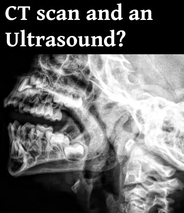
What Is A CT Scan?
A CT scan is a radiological investigation that utilises X-rays to produce images of the internal organs and gives a much more detailed image of bones, muscles, blood vessels, and various organs.
The X-rays are beamed from different directions and different levels, and this helps in the formation of various views of the same organ with much more detail. The image collected at the X-ray plate is sent to a computer where the software produces a 2D image of the structure under study. With the advancement in technology, 3D images are also available now.
It can be done with contrast (to visualise blood vessels better), or without contrast (which is done regularly). Some people may be allergic to this dye, and the severity of the hypersensitivity can vary from person to person. It is important to pass on this information to the doctor if you’ve had a prior reaction to any of the dyes,
A CT scan may pose some risk to pregnant women. It is usually only ordered during pregnancy in conditions where they can’t wait until after the delivery to perform the scan, and when it’s essential in addition to USG and MRI. The treating doctor or the radiologist will make the patient aware of the associated risks if any. Although, the dose of the X-rays used for CT scans is adjusted to a much lower value for pregnant women, and when necessary, CT scans can be done during pregnancy.
Where Is A CT Scan Used?
CT scans can be done in a wide number of cases as well. It’s one of the very commonly used imaging modalities in the emergency department, as it gives a very detailed image of the internal organs and takes much less time to scan the body when compared to an MRI.
A CT scan can be done for:
- Cases of head injury. Various types of intracranial haemorrhage can be visualised in a CT scan.
- Stroke
- Seizures
- Brain tumour
- Chronic headache
- Hydrocephalus
- Congenital intracranial conditions
- Ear, nose, and throat conditions
- Vascular malformations
- Ocular and orbital lesions
- Facial fractures
- Spine injury
- Renal stones
- COVID 19
- To look for lung diseases
- To look for abdominal diseases
- Abdominal injuries and haemorrhages
- To look for metastasis
- Like USG and MRI, it can also be used to perform various guided procedures, like biopsies.
These are some of the conditions where your doctor may ask for a CT scan.
So, What You Need To Remember About a USG and a CT scan?
CT scan and ultrasound are two different types of modalities used for medical diagnoses. USG uses high-frequency sound waves to create an image, while CT scan uses X-rays to produce the image of internal organs.
CT scan gives a more detailed image when compared to a USG, but both have their own set of applications. Even though a CT scan gives a superior image, it’s not required to be done in many cases— where a USG image is enough to make a diagnosis.
Here is a useful video that shows more details about CT scans:
To learn more about radiological imaging, follow this link: Medical Imaging Vs Radiology: What’s The Difference?
Recent Posts
Mastering point cloud to 3d model conversion can feel like translating whispers from another dimension into vivid sculptures. You've got this cloud of data points, a chaotic concert of coordinates...
Let's say you've got a drawing, something you sketched out during a burst of inspiration, and now you're itching to see it leap off the page into three dimensions. Well, that’s exactly what I did...