
An ultrasound anatomy scan is a vital tool in the field of prenatal healthcare. Noninvasive ultrasound imaging enables medical personnel to examine the development of a fetus’ key organs, amniotic fluid levels, and more.
As we look deeper, you will gain insights into the role of ultrasound technicians and diagnostic medical sonographers. These professionals act as crucial patient-physician liaison provider services, bridging gaps between expectant parents and their doctors by providing essential diagnostic information needed for comprehensive prenatal care.
We’ll also explore the timing and preparation required for a routine fetal anatomic survey. Additionally, key features examined during an ultrasound anatomy scan such as monitoring amniotic fluid levels and assessing baby’s heart rate will be discussed in detail.
Finally, I will cover how early identification of potential anomalies like cleft lip can make significant differences in treatment plans. Furthermore, we’ll touch upon maternal health assessment aspects such as evaluating placenta position and checking umbilical cord structure during an anatomy scan.
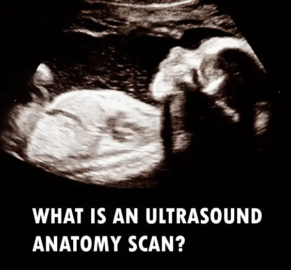
What is the Ultrasound Anatomy Scan
Welcome to the fascinating world of ultrasound anatomy scans.
This procedure, also known as a routine fetal anatomy ultrasound, is a must-have for all expecting parents.
What’s the Scoop?
Anatomy scans are like a backstage pass to your baby’s development. They provide detailed images of your little one’s internal organs and structures in the womb.
A Peek Inside the Womb: What Does an Anatomy Scan Show?
- Fetal heart rate
- Major organs like the brain and spine
- Amniotic fluid level
These scans are like detectives, helping to spot any potential anomalies early on, such as congenital heart defects or cleft lip.
Monitoring amniotic fluid levels during these scans is also crucial for ensuring your baby’s healthy development.
Now that you know what an anatomy scan entails, let’s dive into the preparations and the professionals who conduct these procedures.
Role of Ultrasound Technicians and Diagnostic Medical Sonographers
Let’s first talk about the professionals involved- ultrasound technicians and diagnostic medical sonographers.
The Patient-Physician Liaison Provider Services
An important part of their job? Being the middlemen (or middlewomen) between patients and physicians.
They provide all the diagnostic info needed, from baby’s internal organs to amniotic fluid levels, keeping everyone in the loop.
This info is crucial for getting the right medical care and keeping both mom and baby healthy.
Making Patients Comfortable on The Exam Table
Not only do they measure like pros during a routine fetal anatomic survey, but they also make sure patients are comfy on the exam table.
Liaising with Other Healthcare Providers
In this role, they work closely with insurance medical records visitors maps teams too.
Sonographer’s Responsibility: Providing Essential Info:
Last but not least – Their main gig? Providing all the essential info.
Timing and Preparation for a Routine Fetal Anatomic Survey
The best time for a routine fetal anatomic survey, also known as a level 2 ultrasound, is between 18 to 22 weeks of pregnancy. It’s like a sneak peek into your baby’s world.
This noninvasive prenatal testing gives the medical team all the juicy diagnostic information they need about your little one’s health. No secrets here.
Tips on Preparing For Your Anatomy Scan
To make sure those ultrasound images are crystal clear, here are some things you can do to get ready:
- Hydrate: Guzzle that H2O. Drinking plenty of water before your appointment helps get better baby pics by filling up your bladder and pushing the uterus into a better position. Say cheese.
- Dress Comfortably: Get your comfy clothes on. You’ll be lying down on an exam table with some gel on your belly, so loose-fitting attire is the way to go. Fashion meets function.
- Maintain Regular Check-ups: Keep those appointments coming. Stay on top of your regular check-ups to make sure that any needed interventions or changes can be done quickly. Stay in the loop.
You might be wondering what happens next after all this prep work. Well, get ready for the grand reveal.
In our upcoming section, we’ll dive deep into the fascinating world within the womb and explore the key features examined during an Ultrasound Anatomy Scan.
Key Anatomy Features Examined During an Ultrasound Scan
During an ultrasound scan, the technician closely examines key anatomy features such as the baby’s brain, heart and spine. Talk about an inside job.
Monitoring Amniotic Fluid Levels
Amniotic fluid level plays a crucial role in your little one’s development. It’s like the perfect amount of water in a swimming pool – not too much, not too little. Gotta make sure there’s enough room for those tiny muscles and bones to grow.
Assessing Baby’s Heart Rate
Fetal heart rate monitoring is a big deal during an anatomy scan. We’re talking about the baby’s ticker here. A normal range is usually between 120-180 beats per minute. Anything outside that range might need some extra attention from the medical team.
Identifying Potential Anomalies Early On
The majority of fetal cysts disappear by the 28th week of pregnancy with no effect on the baby.
However, certain anomalies like congenital heart defects need immediate attention because they’re a leading cause of birth defects and infant death.
Recognizing Cleft Lip Early
Cleft lip affects about one in every 600 newborns in the US, making it the fourth most common birth defect.
Now let’s talk about how an anatomy scan can also be crucial for assessing maternal health.
Maternal Health Assessment During Anatomy Scan
An anatomy scan isn’t just about the baby. It’s also a critical tool for assessing maternal health, because let’s face it, moms matter too.
Evaluating Placenta Position
The placenta is your baby’s lifeline during pregnancy, supplying essential nutrients and oxygen. According to Mayo Clinic, its position can influence delivery plans – it should ideally be away from the cervix to allow safe passage for the baby at birth time. So let’s keep that placenta in the right place.
Checking Umbilical Cord Structure
A well-functioning umbilical cord ensures efficient exchange of vital substances between mother and fetus. Ultrasound technicians closely examine potential issues like single artery or knots, because we don’t want any cord complications later on. Safety first.
Remember: an anatomy scan serves dual purposes. Besides giving you glimpses of your little one kicking around inside there (the best part), it provides invaluable diagnostic information needed by the medical team to ensure both mom-to-be’s wellbeing along with her growing bundle of joy.
Records: Navigating the Maze
Medical records can be as confusing as a corn maze.
But fear not, they’re essential for getting top-notch care during your pregnancy.
The Power of Accurate Record Keeping
Keeping your insurance medical records in order is key to getting the best healthcare.
Research shows that clear documentation helps doctors communicate effectively.
Cracking the Code: Understanding Diagnostic Info for Prenatal Care
- Keep track of your routine fetal anatomy ultrasound reports – they’re packed with important diagnostic info for your doctor.
- Well-documented history helps doctors make informed diagnoses, leading to better prenatal care quality.
Unraveling the Importance of Key Statistics and Facts
In the world of ultrasound anatomy scans, numbers speak volumes.
The CDC says cleft lip affects 1 in 600 newborns.
The Significance of Fetal Heart Rate
Fetal heart rate can range from 120-180 beats per minute during an anatomy scan. It’s like a tiny DJ spinning some sick beats.
This helps ultrasound technicians keep tabs on baby’s health. They’re like the medical DJs of the womb.
Anomalies Detected Early On: Congenital Heart Defects
Congenital heart defects are a leading cause of infant death worldwide, according to the World Heart Federation. Let’s give those little hearts some extra love.
FAQs in Relation to Ultrasound Anatomy Scan
What is the importance of an anatomy ultrasound?
An anatomy ultrasound is like a sneak peek into your baby’s world, checking out their major organs and making sure everything is A-OK.
When should you schedule a routine fetal anatomy ultrasound?
For the best view of your little one’s insides, aim for the sweet spot between 18 to 22 weeks of pregnancy.
What’s the deal with an anatomy scan?
An anatomy scan is like a superhero ultrasound, giving you all the diagnostic information your medical team needs to keep your baby healthy.
Is an anatomy scan the same as a regular ultrasound?
An anatomy scan is like a VIP ultrasound, focusing on your baby’s internal organs and other important stuff.
Conclusion
Performed by ultrasound technicians and diagnostic medical sonographers, this noninvasive prenatal testing is the real MVP when it comes to spotting any potential issues early on. From checking the baby’s amniotic fluid levels to evaluating the position of the placenta and umbilical cord, this ultrasound is like a superhero with x-ray vision. It’s like the baby’s own personal exam table, where we can take a closer look at their little heart, making sure it’s beating strong and steady. And if there are any soft markers or signs of congenital heart defects, this ultrasound will catch them faster than a speeding bullet. So, if you’re expecting, don’t skip the routine fetal anatomy ultrasound – it’s the key to getting the diagnostic information needed for proper medical care. Click the following link to learn why you would need a CT scan and an ultrasound.
Recent Posts
Mastering point cloud to 3d model conversion can feel like translating whispers from another dimension into vivid sculptures. You've got this cloud of data points, a chaotic concert of coordinates...
Let's say you've got a drawing, something you sketched out during a burst of inspiration, and now you're itching to see it leap off the page into three dimensions. Well, that’s exactly what I did...