
We have all seen the amazing 3D images of biological specimens displayed, but how are they produced and what microscopes are capable of creating such 3D imagery?
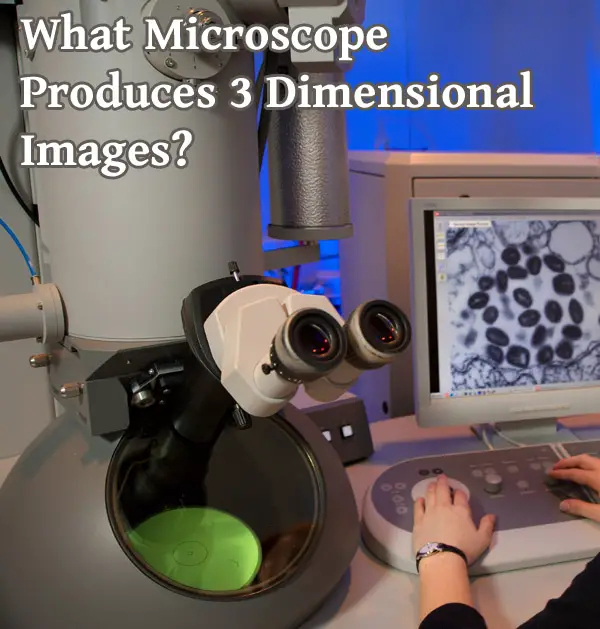
In this article I will discuss the techniques and technology used to produce microscopic 3D images and which types of microscopes can perform this task.
In general, microscopes that produce 3D images include dissecting microscopes, confocal microscopes, and scanning electron microscopes. Other types including transmission electron microscopes and light microscopes may output to 3D utilizing extra steps. 3D images such as volume or surface renderings may be produced in realtime or through the process of 3D reconstruction.
Microscopy has opened the doors to a whole new world of discovery. Prior to the invention of the first microscopes, most information about the unseen, microscopic world was merely theoretical at best. With microscopy, we were now able to get a closer look at these highly impactful organisms and gain more information. However, these microscopic techniques have some limitations.
While the more common light microscopy was able to illuminate the microscopic world, it came at a cost: 2-dimensional (2-D) imaging. Most of the samples had to be flattened in order to allow the light to pass through them in order to create a clear image. Specifically in cytological (cells) or histological (tissue samples), the sample size has to be super thin before being placed between two glass slides in order to be seen due to light penetrance and other issues.
Then on top of that, for even better resolution the investigator would use oil immersion. A drop of specialized oil is placed on the slide to reduce the air between the actual slide and the lens of the microscope. All of that is used to generate a 2-D image.
For 3-dimensional (3-D) images, there has to be a bit more work involved. At one point it was up to the investigator’s imagination to generate these images and simply draw them to the best of their ability. With the use of electron microscopy, we have also been able to view the 3-D surface of cells. As technology has evolved, more has been achieved to go from 2-D to 3-D including developing microscopy that generates real time 3-D imaging.
What do we mean by 3D?
There may be some confusion as to what is actually meant by 3D. 3D refers to viewing something in three dimensions- X, Y and Z and usually being able to rotate the scene, such as with a model of a 3D cell.
This can be achieved by various methods, including viewing in 3D in microscope, realtime 3D rendering, non realtime volumetric or surface rendering, or viewing and manipulating a virtual slide.
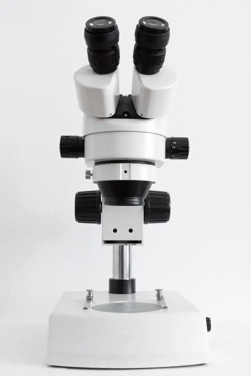
Stereo microscopes- Real Time 3D Viewing
The stereo microscope has been around since the late 1600’s. While not as refined as the ones today, it was still a valuable piece of equipment in generating 3D images in real time without as much of the preparation as the light microscopes and the later electron microscopes. Most people today know of these types of microscopes as dissecting microscopes.
The magnification for these microscopes is considerably low. Where compound light microscopes can reach magnifications into the thousands and electron microscopes into the millions, stereo microscopes can only achieve a magnification of about 10x-50x. This is still greater than an actual magnifying glass (2x-3x). This is because the light is directly reflected from the specimen. The lights on the microscope are used to either illuminate the surface of the specimen (top light) and in cases of more translucent specimens illuminate structures within the specimen (bottom light).
The stereo microscope allows for the investigator to look at a specimen that is normally seen with the naked eye. The image that shows due to the fact that two separate optical paths are utilized including two objectives appears three dimensional, as if you are looking at an object in all its three dimensional reality under an extremely powerful magnifying glass. The reason things appear 3D is light does not go through a flattened specimen on a slide but rather an intact thick specimen lit from all sides. Stereoscopy therefore allows for the illusion of a 3D image.
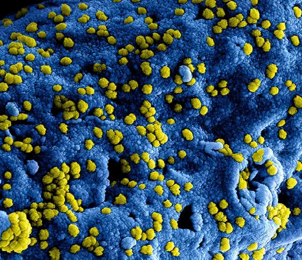
Scanning Electron Microscopy (SEM): 3-D Images
Scanning electron microscopy (SEM) is one of two types of electron microscopy. What separates electron microscopy from light microscopy is the source used to visualize the image. In light microscopy the source is actually light. The light can have different wavelengths that will produce different variations to the image, but the concept is the same.
The light is passed through the sample. The refracted, or bent, beams pass through a one (simple light microscopy) or more commonly a series of lenses (compound light microscopy). The lenses are what allow for resolution (clarity) and magnification (making the image appear larger) of the image.
Electron microscopy uses a different source: electrons. A beam releases a stream of electrons at the sample. The electrons penetrate the sample. The sample then reflects secondary electrons and X-rays that are then detected to generate an image. This is all done in a controlled environment with very expensive machines.
Scanning electron microscopy is used to take images of the surface of the cells. The scanning electrons are meant to penetrate the surface of the cells slightly in order to produce an image. Many of the images seen include structures such as the cell membrane, cell surface proteins, motility structures like cilia or flagella, and other external parts of the cytoskeleton.
The result is a 3D image of extremely small objects which cannot be seen with any other type of microscope. Even though the image appears to be 100% 3D, it cannot be measured as there is no depth information. SEM images can be converted into true 3D objects through the process of 3d surface reconstruction using the data from the SEM image if you want to rotate them.
Because SEM is used to take images of the surface of the cells. The cells have to remain intact. That means these images represent true 3-D images of the cells under examination. Unlike other forms of microscopy, these super thin samples are actually counterproductive since we want to see the intact structures of the cells. The sample sizes could be virtually any size and still have an image generated. The magnification is not as large as transmission electron microscopy, but it is still very impressive and gives a more accurate representation of what happens with the cell membrane and/or cell wall.
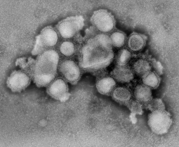
Transmission electron TEM- Through 3D Surface Rendering
On the other hand, transmission electron microscopy (TEM) requires a bit more work in order to achieve a 3D rendering. TEM requires a more involved sample preparation before the microscopy can even be performed. The sample must be thin so an ultramicrotome is called for to produce a sample that is less than 150 nm in thickness.
Transmission electron microscopy is also more specialized and requires trained professionals in order to use them. Once the sample is prepared and placed, transmitted electrons are passed through the sample. The electrons that get through the sample are then collected in a lens below the sample and an image is generated. Picture a stencil where you place different colors of sand on top. Once you lift the stencil, you are given an image.
In order to generate a 3D image with TEM, serial sections need to be photographed and from there a 3d model can be created based on the images in the stack . The basic concept is that multiple images are taken of multiple sections of the samples. From there computer software is used to generate a 3D image of the cells and corresponding structures. This process does take time and additional labor, but has been shown to be fairly successful.
Confocal Microscopy – 3D Surface or Volume Rendering
Confocal microscopy is part of the spectrum of fluorescence microscopy. It is used to give more detailed and defined images of cellular structures at different depths within the cells. Unlike light microscopy and electron microscopy, live specimens can be used and are typically still viable after the process is completed with minimal light damage.
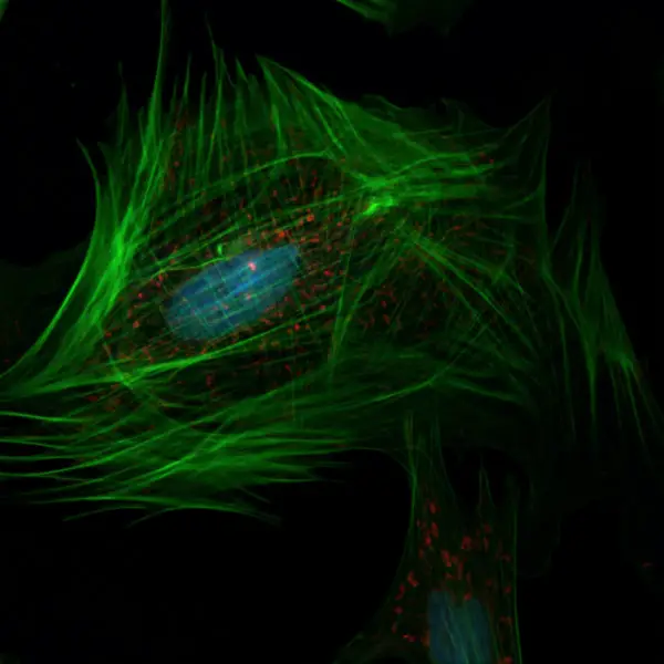
Confocal microscopy uses fluorescence optics but the process is more refined. Instead of releasing a widespread light over the specimen and using the refracted light to generate the best image possible, a very small focused stream of light is directed through the sample at a very specific depth. The light or point of illumination is then reflected back to the lens without the remaining unfocused light yielding a very small yet highly focused image. This process is repeated throughout the area of the sample to at the same depth to reveal a 2D image with higher resolution.
In order to generate a 3D image, this process must be done multiple times at different depths to generate many 2D images in a “3d stack”. Those images are then combined to generate a 3D rendering of the specimen. Such a rendering can be a volume rendering or a surface (polygon) rendering.
With newer confocal microscopes, however, it is now possible to render the image on the fly in 3D in the included microscope software. The future seems very bright for 3D in microscopy.
Virtual Slides
There is another form of 3D microscopic imaging that requires extra work- virtual slides (whole slides). These are scans of an actual specimen slide taken at different depths and magnifications to give you a digital version of a physical slide which will not degrade over time and can be easily transmitted from computer to computer. Virtual slides can be viewed using online or software based viewers.
Here is an interesting video on how a scanning electron microscope works:
Conclusion
As microscopy has grown the technical aspects of generating high resolution images with detailed impressions of structures has become an even more accessible reality. With the application of software technology, a lot of those super detailed while flat images can be utilized to generate 3D renderings of microscopic specimen samples. In some cases, those images can even be taken of live specimens while still leaving them viable for use in other experiments.
As the technology around microscopy grows, perhaps it will come to meet the ingenuity of those that utilize it.
Click the following link to learn what hardware you need for 3D rendering.
Recent Posts
Mastering point cloud to 3d model conversion can feel like translating whispers from another dimension into vivid sculptures. You've got this cloud of data points, a chaotic concert of coordinates...
Let's say you've got a drawing, something you sketched out during a burst of inspiration, and now you're itching to see it leap off the page into three dimensions. Well, that’s exactly what I did...