
Are you knee deep in microscope slides examining the morphology of different cells? Are you just seeing a bunch of pretty shapes and colors without a full grasp on what the circles and squiggles do? A box of microscope slides represents a huge cellular graveyard. While helpful, sometimes motion is needed to get the full picture.
In this article I will explain whether fluorescence microscopy kills cells and explain the reasoning.
Fluorescence microscopy does not kill cells and does not require the killing of cells to achieve an image. The dyes that are utilized in the process are not toxic to the cells. In addition to that, the cells are not required to be fixed or stationary in order for the images to be taken. In fact, fluorescence microscopy allows for a researcher to view live cells and take images in real time to examine changes within the cells. Those images can be combined to create a “movie” effect allowing a full understanding of what is happening within specific cells.
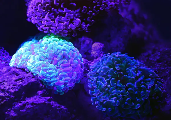
There are many organisms that can be visualized through fluorescence. Many of our known marine life that live deep within the ocean have some sort of fluorescent ability that allows them to attract prey, drive away predators, and/or send signals to other members of their group. Many of these organisms fluoresce through symbiotic relationships of microorganisms. The same methodologies are applied in the realm of fluorescence microscopy. The dyes are not harmful to the cells that are tagged, and the dyes allow the cells and their structures to be easier to visualize under a microscope.
The Need for Fluorescence Microscopy
As cell and molecular biology become more of the standard in biological study, advances in microscopy are going to be an ever present demand. Unfortunately, cells in real life do not look as photogenic nor as beautiful as the images we draw in textbooks. Cells under a microscope are typically colorless unless they are cells containing pigment vesicles. Even in those cases, the vesicles are the only pigment containing portions or organelles.
Our solution is to look at different types of dyes in order to stain certain organelles without impacting other organelles. This is the foundation behind the Gram stain which is our most basic diagnostic technique. The staining of a cell wall in a bacterium allows us to not only see the bacterium under a microscope but allows medical professionals to diagnose a disease and treat.
When it comes to research, the biggest hindrance when it comes to cellular processes is fixation. Fixation is the process by which a cell is adhered to a surface, typically a microscope slide or the bottom of a Petri dish or flask. Some cells do this naturally. Somatic cells for instance are studied and grown both in solution (floating around in the culture medium) as well as fixed depending on what is needed for the study.
Chemical and heat fixation of cells before examination under a microscope automatically kills the cells allowing us to examine the structures unhindered by movement and capture a static image. Nevertheless, when it comes to seeing processes a dead cell doesn’t help as much. You can capture many images of stages of cell division, but to be able to see the process from start to finish, the cells need to be alive. One major solution is fluorescence microscopy.
Fluorescence microscopy is another piece of the puzzle when it comes to examining cellular life and processes. Where cellular fixation can freeze a cell in a moment and examine what happened right as the cell was fixed, fluorescence microscopy can freeze an infinite amount of moments by taking multiple pictures of a cell. The fact that this can be used in live cells means that we are no longer in the dark as to how the cell arrived at a specific position. Thanks to multiple images, we can compile them chronologically and watch how a cells organelles maneuver with the cell membrane and see exactly what is changing and how fast it is changing. You can watch an entire round of mitosis without every having to fix a cell!
Fluorescence Microscopy: In a Nutshell
Fluorescence Microscopy is usually one of the middle tier steps when it comes to doing research. At this stage, a lot of the preliminary exploratory experiments are done. Now, the publishable results can begin. Some of the most beautiful images are taken with microscopy. These images have a way of bringing a research presentation to life. Whether it is examining how a drug impacts certain cellular processes or just observing some new cellular phenomenon, the point is that these things need to be seen and photographed.
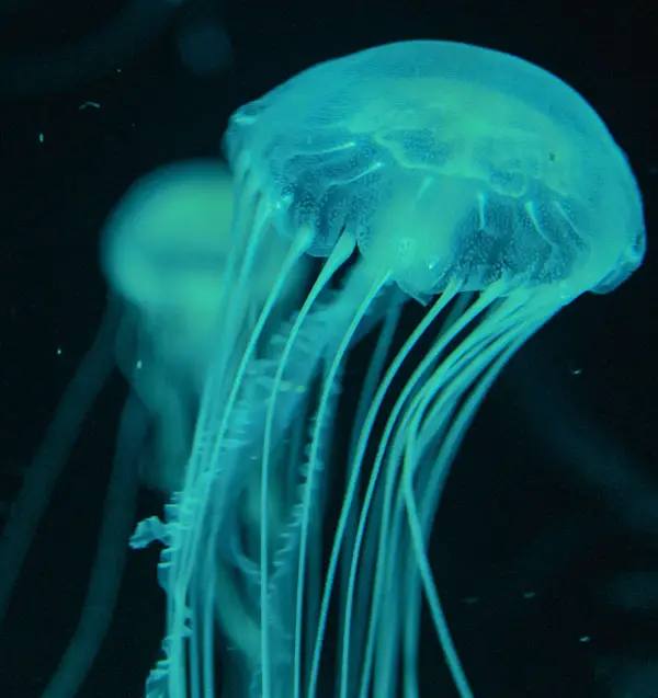
For the most part, an individual cell is fairly transparent. Like the jellyfish, we cannot see them without assistance unless they are dead. The basics of fluorescence microscopy is the addition of a certain dye to the different organelles of the cell. Depending on which organelles you want to see, multiple dyes are necessary. The mitochondria may need to be one color, the nucleus another, and the differing filaments of the cytoskeleton will be other colors. The live sample is then placed under a microscope with different emission and excitation filters that are specific to the types of dyes used.
The excitation filters are used to filter the lights emitted from the light source. This is usually a shorter wavelength and thereby brighter light that may be more difficult to see. An excitation filter will help diffuse that light more and allow the light to make contact with the entire surface of the sample. The emission filter comes into effect by allowing certain light to come in contact with the sample. This then allows certain organelles to emit light. All of these filters are arranged in a cube and swapped out in accordance to what needs to be seen.
A great example of this is the blue light that is emitted from our handheld devices. The light shines into our eyes and causes eye strain. This can result in dryness as well as fatigue. The screen on your tablet or phone is the excitation filter because it allows the light to be released from the device. Without an emission filter, the blue light will shine directly onto the sample and directly into the eyepiece and into the eye. The solution to this is blue light lenses. Blue light lenses do not permit blue light to enter into the eye directly. The same goes for an emission filter. Without an emission filter, the light would be too bright to allow for visualization of the cells. An emission filter disperses the light and only allows certain light colors through. The colors that are allowed correspond to the colors that were used as fluorescent dyes.
Fluorescence Microscopy: Uses and Benefits
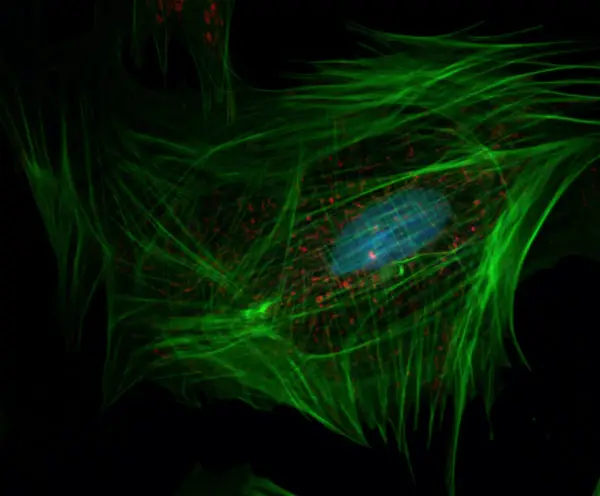
At this point, images of the cells being examined can be taken. The high contrast of the black background to the emitted light allows for these structures to be seen easily and in high resolution. After those images are collected, the filters are changed to illuminate another organelle or cellular structures. The process is repeated again and again until all of the images are collected. These images are then layered on top of one another to illustrate all of the different organelles as they would appear within a cell. The images can then be organized chronologically to show the cellular processes in motion.
Dr. Nico Sturrman of the University of California San Francisco and the Howard Hughes Medical Institute has an amazing video introducing the viewers to the concepts of fluorescence microscopy including theory and application. He also shows some of his own images taken of live cells and how he combined them to show different structures and create a “movie” of a cell dividing:
The Necessity of Microscopic Graveyards?
As we continue our study of life, even at the microscopic level, these images are just another piece of the puzzle. While it is great to see how these cells move in live action, there is still a need to look at structures from a static perspective. That is the purpose of other forms of microscopy that require cellular fixation and other manipulations. For example, electron microscopy requires fixation along with slicing of the cells and many other steps depending on which type is required (TEM vs. SEM).
Fluorescence microscopy is probably one of the more attractive ways to view cells and study them over time. Much like the animals we discover in the ocean, the brighter, colorful ones tend to be the most attractive. The angler fish uses that technique very successfully. Anyway, fluorescence microscopy is a great way to examine cells prior to fixation and allows for more information to be added to create more of a completed picture for research.
Check out our article on the maximum magnification of a confocal microscope.
Recent Posts
Mastering point cloud to 3d model conversion can feel like translating whispers from another dimension into vivid sculptures. You've got this cloud of data points, a chaotic concert of coordinates...
Let's say you've got a drawing, something you sketched out during a burst of inspiration, and now you're itching to see it leap off the page into three dimensions. Well, that’s exactly what I did...