
Microscopes have come a long way since they were first created. From a simple compound microscope to an electron and atomic force microscope—there are a variety of microscopes available to choose from, depending on the purpose. We can see our own cells or other microscopic organisms and their components under a microscope.
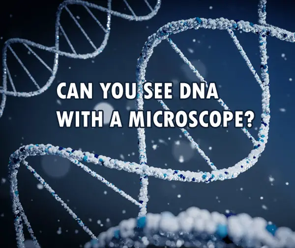
But can we see DNA using a microscope?
DNA cannot be seen in the regular compound microscope. But DNA can be seen with the help of more advanced methods like electron microscopy and atomic force microscopy. You can see the nucleus with a compound microscope, but not the DNA. Chromosomes can be seen only during cell divisions.
Compound microscopy is great to see a cell, its components and various organisms. Details of the various organelles can be seen with the maximum magnification of a compound microscope.
Nucleus and chromatin can be easily seen with a compound microscope.
Chromosomes can be seen only during cell division. They are first seen during the prophase of cell division. Chromosomes can be visualised at 40X magnification.
However, DNA is very small and therefore cannot be seen with a regular compound microscope. To see DNA, you need more advanced techniques like electron microscopy.
What Kind Of Microscope Can Be Used To See DNA?
At present, DNA can be best visualised with an electron microscope.
DNA was discovered in 1952 based on the X-ray diffraction image, known as photo 51. It was more of an indirect way of looking at the DNA and concluding that it is helical in shape. The conclusion was based on the X-pattern produced on the X-ray diffraction and the mathematical understanding that a helical structure would produce an X-pattern on X-ray diffraction. The DNA helix itself was not directly seen.
It wasn’t until very recently that we actually had the images of the DNA helix. In 2012, using an electron microscope, the image of the DNA helix was captured for the first time. It was captured by stretching the DNA strands between silicone nanopillars and then firing a beam of electrons on it to visualise and capture the images of the actual DNA helix.
However, even with electron microscopy, we can’t see a single DNA. What we see with an electron microscope is a “cord” of DNA, containing a minimum of 6 DNA molecules wrapped around a 7th DNA molecule (acting as a base). This is because a single can’t withstand the energy produced by the beam of electrons.
Another method that has been used to see the DNA is atomic force microscopy(AFM).
AFM uses scanning probes to see the images. The probe touches and “feels” what the structure looks like and based on that an image is produced. It doesn’t use any lenses or beam radiation to produce an image.
Can You See Chromosomes And Genes With A Microscope?
The chromosomes, genes and DNA are present within the nucleus of a cell. The chromosome contains the DNA along with other proteins. On the other hand, genes are the segments of DNA. It is the difference in the molecular sequence of these segments (i.e. genes) that give us our individual characteristics.
Chromosomes can be seen with a compound microscope only when the cell is dividing. This is because during cell division the chromosomes condense at a certain place with the nucleus, making it easier to see the collection of chromosomes.
Chromosomes can be first seen on a light microscope during the leptotene stage of the prophase. This is when the chromosome begins to condense at a point.
The best microscope to see a chromosome, however, would again be with electron microscopy, like a scanning electron microscope.
Genes are studied by studying the sequence of the nucleotides in the DNA. To study a gene, various molecular methods are used. Very commonly fluorescence microscopy and FISH are used to see the gene sequence. Gene is a small part of DNA, and therefore cannot be seen with a compound microscope.
DNA, as mentioned earlier, can be seen with an electron microscope, and atomic force microscopy has also been tried to see the DNA helix.
Methods To Check The DNA Yield In The Sample
Once the sample is collected for DNA visualisation or testing, to check whether the sample has a good amount of DNA or not certain quantification methods can be used.
Agarose gel electrophoresis is one such technique using which the DNA in the sample can be separated from the proteins using agar and electrodes. On passing the current, as DNA is negatively charged, it moves towards the positive electrodes. This way we can see the quantity of DNA in the sample, as well as separate the DNA from other proteins.
Fluorescent DNA-binding dyes can also be used to visualise DNA fragments, and when shown UV light, they fluoresce, making it easier to see the presence of DNA.
UV absorbance is another method to quantify DNA. The sample is exposed to 260nm of UV light. This is because the nucleic acids have maximum absorbance at 260nm wavelength. However, this isn’t the best method for DNA quantification as it only works for pure DNA samples. The presence of other proteins can interfere with the results.
So, these are some common ways to check for the yield of DNA in a sample. The separated DNA can then be used for various purposes— to visualise under a microscope, DNA fingerprinting, and other molecular studies.
What To Remember About Visualising DNA Under A Microscope?
DNA is very small in size and therefore it cannot be seen with a light microscope. However, they can be visualised using more advanced microscopes like electron microscopes and atomic force microscopes.
Chromosomes are best seen under electron microscopes as well. But they can also be seen with a compound microscope during certain phases of cell division.
Genes, on the other hand, can be studied with the help of various molecular methods like FISH to identify the location of a particular gene on the chromosome.
You can read about the types of microscopes that produce 3D images here.
Recent Posts
Mastering point cloud to 3d model conversion can feel like translating whispers from another dimension into vivid sculptures. You've got this cloud of data points, a chaotic concert of coordinates...
Let's say you've got a drawing, something you sketched out during a burst of inspiration, and now you're itching to see it leap off the page into three dimensions. Well, that’s exactly what I did...