
Why is 3D reconstruction not done for every CT scan or MRI? This is a question that often arises in the domain of medical imaging technology.
After all, 3D reconstructions provide the most life like depiction of anatomy and pathology available, while other modalities only show sectional 2D views
The answer, like most things in medicine and tech, isn’t as straightforward as one might hope.
In fact, the use of 3D reconstructions from CT scans or MRIs can be quite complex. The process involves intricate software tools and requires significant time investment.
This complexity may deter some from utilizing it consistently. Yet understanding why 3D reconstruction is not always used could offer valuable insights into its potential benefits and limitations.
The Evolution of 3D Reconstruction in Medical Imaging
Over the past decades, medical imaging has seen remarkable advances with 3D reconstruction from CT scans and MRIs leading to improved operative planning and patient outcomes. The advent and refinement of 3D reconstruction from CT scans and MRIs have brought about revolutionary changes to how clinicians approach operative planning and enhance patient outcomes. However, these techniques are not employed universally for every scan due to several constraints.
One primary hurdle is cost-related. Advanced software tools required for generating high-quality three-dimensional image simulations, especially realtime ones, can be expensive, thus deterring smaller healthcare facilities or those with limited budgets from investing heavily in them. Elarref MA et al. suggests that another challenge lies within the complexity associated with certain reconstructions, such as lung reconstruction or advanced mediastinal mass reconstruction, which require specific expertise often only found among specialized professionals.
Volumetric Data Acquisition: A Paradigm Shift?
In recent years, advancements aimed at making volumetric data acquisition more accessible and efficient have emerged on the horizon. Note: These include open-source medical image viewers like Horos, enabling professionals across different surgical specialties, including general thoracic surgery cases or cardiothoracic surgery specifically, to utilize free 3D modeling software without hefty investments.
This democratization of technology has also resulted in real-time interactive 3D imaging becoming increasingly prevalent within clinical practice around the globe.
Decoding the Process of 3D Reconstruction
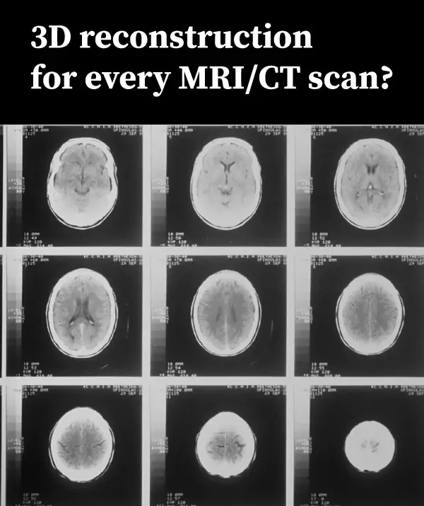
The realm of medical imaging has seen significant advancements with the introduction and evolution of 3D reconstruction. But what exactly does this process entail? Let’s delve into how CT scans or MRIs are transformed from two-dimensional images to three-dimensional representations using advanced software tools.
These scanning techniques capture cross-sectional views, each representing a thin slice of internal body structures at various angles. When these slices come together, they form a volumetric dataset that serves as raw material for creating our desired output – interactive 3D reconstructions.
Leveraging Free Tools for Basic Reconstructions
In an era where resources can be limited, free alternatives have emerged offering valuable assistance to professionals looking to utilize basic reconstruction methods effectively without breaking their budget. Horos, an open-source medical image viewer, offers capabilities such as volume rendering and multiplanar reformation, making it an ideal tool when considering a patient’s CT scan and how to perform tasks like lung reconstructions.
This platform allows users to import DICOM files – a standard format for storing medical imaging data – directly from devices or PACS servers. However, while Horos provides control over visualization tasks, it may lack some features found in commercial counterparts, like sophisticated segmentation capabilities or post-processing filters.
To navigate through these limitations successfully, one must carefully consider specific needs before opting for any particular tool, whether a paid version or a free alternative.
Applications of Advanced Visualization in General Thoracic Surgery
The transformative power of advanced visualization tools has revolutionized general thoracic surgery. By enabling a comprehensive understanding of complex anatomical structures, these technologies have significantly improved surgical planning and patient outcomes.
In the educational context, interactive platforms provided by these visualizations allow for demonstration and exploration of intricate procedures or conditions. Trainees can manipulate 3D models to view different angles and layers that traditional 2D images cannot provide, gaining an all-encompassing perspective on the subject matter at hand.
A Case Study on Operative Planning
One area where advanced visualization truly shines is operative planning. A study comparing imaging techniques, for instance, showcased how CT scans could be utilized in operative planning specifically within cardiothoracic surgery.
This study demonstrated that high-quality three-dimensional image simulation resulted in superior preoperative assessment compared to conventional methods alone, which often lack sufficient detail or spatial context required for complex surgeries such as lung reconstruction or re-do surgery operations.
Surgeons were able to visualize tumors relative to surrounding tissues with this technology before stepping into the operating room, allowing them meticulous preparation leading to potentially better short-term surgical outcomes by reducing complications associated with unexpected intraoperative findings.
Patient Education Benefits from Advanced Visualization
An equally important application lies within patient education – a critical aspect of the healthcare delivery system. With sophisticated modeling software, physicians are now equipped to create personalized simulations based on an individual’s own medical imagery. By leveraging personalized simulations based on an individual’s medical imagery, physicians can enable patients to gain a better comprehension of their condition and the treatment options available, leading to increased satisfaction post-surgery.
The Role of Interactive 3D Reconstruction in Video-Assisted Thoracoscopic Lung Surgery
Video-assisted thoracoscopic surgery (VATS) is a game-changer for lung surgery, offering less invasive procedures that lead to reduced postoperative discomfort and faster recovery. However, VATS demands an intricate understanding of complex pulmonary anatomy, which can be difficult to visualize using traditional two-dimensional imaging techniques.
This is where interactive 3D reconstruction steps in. By converting CT scans into three-dimensional models, surgeons gain deeper insights into the patient’s unique anatomy and plan their surgical approach accordingly.
Paving Way for Preoperative Planning with Interactive 3D Reconstructions
In terms of preoperative planning, these reconstructions allow surgeons to virtually navigate through the operative site before making an incision. They can identify potential obstacles such as tumors or anatomical variations that may complicate the procedure ahead of time.
The ability to rotate and manipulate these models also gives them a comprehensive view from multiple angles – something not possible with conventional imaging methods. This greatly enhances their spatial awareness, leading to more accurate operative plans, specifically enhancing cardiothoracic surgery.
Virtually Simulating Surgical Procedures: A Leap Forward
Beyond just planning purposes, interactive 3D reconstructions offer opportunities for virtual simulation too. Surgeons are now able to rehearse complicated surgeries beforehand on a computer screen, simulating real-life conditions closely.
This practice leads to improved performance during actual surgeries due to its high fidelity nature, accurately mirroring real-world scenarios. This increases surgeon confidence while significantly reducing error rates, according to recent studies, including one conducted by Dr. Nia’s team mentioned earlier in this article.
Unraveling the Challenges and Limitations of Current 3D Reconstruction Techniques
In our journey to understand advanced imaging techniques, it is essential to address not just their advantages but also their limitations. These technologies are intended as adjunct tools rather than replacements for traditional axial imaging.
Precision Matters: The Accuracy Dilemma
The precision of basic mediastinal mass reconstruction heavily depends on the clarity and resolution of initial images from CT scans or MRIs. Any artifacts present could affect subsequent visualizations, potentially leading to misinterpretation.
Software algorithms have improved over time, yet they still struggle with complex anatomical structures where different tissues intersect closely together, such as lung parenchyma or vascular trees.
Lack Of Standardization And Training
Different platforms used for generating interactive 3D reconstructions can produce varying results due to differences in underlying algorithms. This lack of standardization can lead to inconsistencies when comparing studies conducted using various systems.
Apart from this technical aspect, there is an absence of standardized training for medical professionals on how best to utilize free modeling software – an issue that needs addressing if we want wider adoption within healthcare institutions worldwide.
Exploring Open Source Medical Image Viewers
The realm of open source medical image viewers is expansive and diverse, offering a myriad of tools for professionals in the scientific 3D field. These platforms present an economical alternative to expensive proprietary software, making advanced reconstruction more attainable.
RadiAnt DICOM Viewer, with its user-friendly interface and comprehensive functionality, has become quite popular. It supports various modalities such as CT scans, MRIs, PETs, and ultrasounds, among others. The unique feature it provides – a 3D cursor that synchronizes points picked across different series – enables easier comparison between images.
Moving beyond basic viewing functionalities offered by RadiAnt lies 3D Slicer, an interactive platform designed specifically for medical image processing, particularly useful for research purposes. From simple measurements or visualizations to complex surgical simulations or radiotherapy planning procedures, they can be performed using this powerful tool.
This robust tool facilitates extensive options for segmentation, where regions within the anatomy can be separately highlighted, allowing detailed study of structures inside a patient’s body based on their CT scan. How-to instructions provided by radiology experts further enhance the utilization of this tool.
Looking Towards the Future – Augmented Reality Visualization
The future of 3D imaging, particularly in thoracic surgery and other surgical specialties, holds immense promise with augmented reality (AR) visualization. This cutting-edge technology is set to redefine patient outcomes by enhancing surgical precision.
Augmented reality overlays digital data onto real-world environments, providing surgeons with a comprehensive view of complex anatomical structures without making invasive incisions. It is poised to reduce operative times while simultaneously improving accuracy.
Potential Advantages of AR
AR offers an enhanced understanding and interactive 3D reconstruction for intricate anatomy like lung tissues. These visual aids are instrumental for effective preoperative planning and intraoperative guidance during procedures such as lung resections or advanced reconstructions.
A study led by Elarref MA et al. underscores how AR visualization can lead to more precise tumor resection margins along with improved lymph node dissection techniques – potentially optimizing short-term surgical outcomes for patients undergoing these types of surgeries.
Leveraging Open Source Platforms For Implementing AR
To realize widespread adoption of this innovative technique, it will be crucial to utilize open-source software platforms, allowing professionals easy access to high-quality simulations without hefty investments.
Developers have already started creating open-source projects aimed at implementing augmented reality into healthcare settings – indicating promising signs towards a future where every surgeon has ready access to interactive 3D reconstruction tools coupled with augmented reality capabilities.
As we forge ahead into this exciting new era in medicine, technologies like AR undoubtedly play pivotal roles in shaping our approach towards achieving optimal results across various surgical specialties.
FAQs
Can CT scans produce 3D images?
Yes, with the help of advanced software tools, data from CT scans can be converted into detailed 3D reconstructions for better visualization and analysis.
What is 3D reconstruction in CT scan?
It’s a process where two-dimensional X-ray images captured during a CT scan are processed using specialized software to create a three-dimensional model of the scanned area.
Why do MRIs have better tissue differentiation compared with CT scanners?
MRI uses magnetic fields and radio waves instead of radiation. This allows it to differentiate between tissues more effectively than a standard CT scanner, providing clearer imaging results.
What are the advantages of using 3D reconstructions?
The use of 3D reconstructions provides enhanced spatial understanding, aids in surgical planning, improves patient education, and has potential applications in teaching medical students or trainees.
Conclusion
Progress in 3D reconstruction for medical imaging has been significant, from being a costly and time-consuming procedure to becoming more attainable with the introduction of sophisticated software solutions. While it’s proving invaluable in areas like thoracic surgery for teaching, patient education, and operative planning, it’s not without its challenges. It serves as an adjunct tool rather than replacing axial imaging entirely.
The potential pitfalls and limitations of current techniques underscore the need for further advancements in this field. This brings us to open source medical image viewers that allow professionals to create high-quality simulations sans hefty investments.
As we look towards the future, augmented reality visualization stands out as a promising trend that could revolutionize surgical specialties beyond just thoracic surgery. Click the following link to learn about the best free DICOM viewers.
Recent Posts
Mastering point cloud to 3d model conversion can feel like translating whispers from another dimension into vivid sculptures. You've got this cloud of data points, a chaotic concert of coordinates...
Let's say you've got a drawing, something you sketched out during a burst of inspiration, and now you're itching to see it leap off the page into three dimensions. Well, that’s exactly what I did...